 Explore
 Validate
 Learn
Learn Western blot
Antibody data
- Antibody Data
- Antigen structure
- References [2]
- Comments [0]
- Validations
- Western blot [4]
- Immunocytochemistry [2]
- Flow cytometry [1]
- Other assay [1]
Submit
Validation data
Reference
Comment
Report error
- Product number
- 701421 - Provider product page

- Provider
- Invitrogen Antibodies
- Product name
- Cyclin D1 Recombinant Rabbit Monoclonal Antibody (17H3L3)
- Antibody type
- Monoclonal
- Antigen
- Synthetic peptide
- Description
- Intact IgG appears on a non-reducing gel as ~150 kDa band and upon reduction generating a ~25 kDa light chain band and a ~50 kDa heavy chain.
- Antibody clone number
- 17H3L3
- Concentration
- 0.5 mg/mL
Submitted references Fusobacterium nucleatum promotes colorectal cancer by inducing Wnt/β-catenin modulator Annexin A1.
CCND1-CDK4-mediated cell cycle progression provides a competitive advantage for human hematopoietic stem cells in vivo.
Rubinstein MR, Baik JE, Lagana SM, Han RP, Raab WJ, Sahoo D, Dalerba P, Wang TC, Han YW
EMBO reports 2019 Apr;20(4)
EMBO reports 2019 Apr;20(4)
CCND1-CDK4-mediated cell cycle progression provides a competitive advantage for human hematopoietic stem cells in vivo.
Mende N, Kuchen EE, Lesche M, Grinenko T, Kokkaliaris KD, Hanenberg H, Lindemann D, Dahl A, Platz A, Höfer T, Calegari F, Waskow C
The Journal of experimental medicine 2015 Jul 27;212(8):1171-83
The Journal of experimental medicine 2015 Jul 27;212(8):1171-83
No comments: Submit comment
Supportive validation
- Submitted by
- Invitrogen Antibodies (provider)
- Main image
- Experimental details
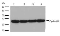
- Western blot analysis of Cyclin D1 in whole cell extracts from U87-MG, A549, MCF-7, and U20S (lanes 1-4 respectively) using a Cyclin D1 recombinant rabbit monoclonal antibody (Product # 701421) at a dilution of 1 µg/mL. Detection was performed using an HRP-conjugated goat anti-rabbit secondary antibody followed by chemiluminescence (ECL). Results show a band at ~36kDa.

- Submitted by
- Invitrogen Antibodies (provider)
- Main image

- Experimental details
- Western blot analysis of Cyclin D1 was performed by loading 30 µg of HeLa, A549, MCF-7 and U-2 OS cell lysates using Novex®NuPAGE®4-12% Bis-Tris gel (Product # NP0321BOX), XCell SureLock Electrophoresis System (Product # EI0002), Novex® Sharp Pre-Stained Protein Standard (Product # LC5800), and iBlot® Dry Blotting System (Product # IB21001). Proteins were transferred to a nitrocellulose membrane and blocked with 5% skim milk for 1 hour at room temperature. Cyclin D1 was detected at ~36 kDa using Cyclin D1 Recombinant Rabbit Monoclonal Antibody (Product # 701421) at a 1:1000 dilution in 2.5% skim milk at 4°C overnight on a rocking platform. Detection was performed using an HRP-conjugated Goat anti-Rabbit secondary antibody (Product # G-21234) at a 1:5000 dilution and chemiluminescent detection was performed using Pierce™ ECL Western blotting Substrate (Product # 32106).

- Submitted by
- Invitrogen Antibodies (provider)
- Main image
- Experimental details
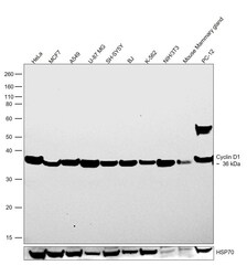
- Western blot was performed using Anti-Cyclin D1 Recombinant Rabbit Monoclonal Antibody (17H3L3) (Product # 701421) and a 36 kDa band corresponding to G1/S-specific cyclin-D1 was observed. Whole cell extracts (30 µg lysate) of HeLa (Lane 1), MCF7 (Lane 2), A549 (Lane 3), U-87 MG (Lane 4), SH-SY5Y (Lane 5), BJ (Lane 6), K-562 (Lane 7), NIH/3T3 (Lane 8), tissue extract (30 µg lysate) of Mouse Mammary gland (Lane 9) and whole cell extract of PC-12 (Lane 10) were electrophoresed using NuPAGE™ 10% Bis-Tris Protein Gel (Product # NP0302BOX). Resolved proteins were then transferred onto a nitrocellulose membrane (Product # IB23001) by iBlot® 2 Dry Blotting System (Product # IB21001). The blot was probed with the primary antibody (1:2000) and detected by chemiluminescence with Goat anti-Rabbit IgG (H+L) Superclonal™ Recombinant Secondary Antibody, HRP (Product # A27036,1:20,000) using the iBright FL 1000 (Product # A32752). Chemiluminescent detection was performed using SuperSignal™ West Pico PLUS Chemiluminescent Substrate (Product # 34580).

- Submitted by
- Invitrogen Antibodies (provider)
- Main image
- Experimental details
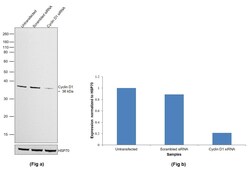
- Knockdown of G1/S-specific cyclin-D1 was achieved by transfecting HeLa with G1/S-specific cyclin-D1 specific siRNAs (Silencer® select Product # S230, S229). Western blot analysis (Fig. a) was performed using Whole cell extracts from the G1/S-specific cyclin-D1 knockdown cells (lane 3), non-targeting scrambled siRNA transfected cells (lane 2) and untransfected cells (lane 1). The blot was probed with Cyclin D1 Recombinant Rabbit Monoclonal Antibody (17H3L3) (Product # 701421, 1:3000 ) and Goat anti-Rabbit IgG (H+L) Superclonal™ Recombinant Secondary Antibody, HRP (Product # A27036, 1:20,000). Densitometric analysis of this western blot is shown in histogram (Fig. b). Decrease in signal upon siRNA mediated knock down confirms that antibody is specific to G1/S-specific cyclin-D1.

Supportive validation
- Submitted by
- Invitrogen Antibodies (provider)
- Main image

- Experimental details
- Immunofluorescent analysis of Cyclin D1 in HeLa cells using a Cyclin D1 recombinant rabbit monoclonal antibody (Product # 701421) followed by detection using an Alexa Fluor 488-conjugated goat anti-rabbit secondary antibody (green) (Image A). Nuclei were stained using DAPI (Image B) and actin stained with Alexa Fluor 594 phalloidin (red) (image C). Image D is a composite image showing cytoplasmic and nuclear localization of Cyclin D1.

- Submitted by
- Invitrogen Antibodies (provider)
- Main image

- Experimental details
- Immunofluorescence analysis of G1/S-specific cyclin-D1 was performed using 70% confluent log phase Hep G2 cells. The cells were fixed with 4% paraformaldehyde for 10 minutes, permeabilized with 0.01% Triton™ X-100 for 15 minutes, and blocked with 2% BSA for 45 minutes at room temperature. The cells were labeled with Cyclin D1 Recombinant Rabbit Monoclonal Antibody (17H3L3) (Product # 701421) at 1:100 in 0.1% BSA, incubated at 4 degree celsius overnight and then labeled with Donkey anti-Rabbit IgG (H+L) Highly Cross-Adsorbed Secondary Antibody, Alexa Fluor Plus 488 (Product # A32790), (1:2000), for 45 minutes at room temperature (Panel a: Green). Nuclei (Panel b:Blue) were stained with Hoechst 33342 (Product # H1399). F-actin (Panel c: Red) was stained with Rhodamine Phalloidin (Product # R415, 1:300). Panel d represents the merged image showing predominant cytoplasmic localization. Panel e represents control cells with no primary antibody to assess background. The images were captured at 40X magnification in CellInsight CX7 LZR High-Content Screening (HCS) Platform (Product # CX7C1115LZR).

Supportive validation
- Submitted by
- Invitrogen Antibodies (provider)
- Main image

- Experimental details
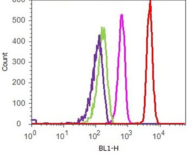
- Flow cytometry analysis of Cyclin D1 was performed on serum-starved HeLa cells. Cells were fixed with 70% ethanol for 10 minutes, permeabilized with 0. 25% Tritonª X-100 for 20 minutes, and blocked with 5% BSA for 1 hour at room temperature. Cells were labeled with ABfinityª Cyclin D1 recombinant rabbit monoclonal antibody (Product # 701421, red histogram) or with rabbit isotype control (pink histogram) at a dilution of 1:400 in 2.5% BSA. After incubation at room temperature for 3 hours, the cells were labeled with Alexa Fluor¨ 488 goat anti-Rabbit Secondary antibody (Product # A11008) at a dilution of 1:400 for 30 minutes at room temperature. The representative 10,000 cells were acquired and analyzed for each sample using an Attune¨ Acoustic Focusing Cytometer. The purple histogram represents unstained control cells and the green histogram represents no-primary-antibody control.
Supportive validation
- Submitted by
- Invitrogen Antibodies (provider)
- Main image

- Experimental details
- Figure 1. Elevated CCND1-CDK4 (4D) expression induces a G0-to-G1 transition and increased signaling in human HSPCs. (A) Scheme of the construct: CCND1 and CDK4 are linked by T2A, followed by an IRES-GFP-reporter sequence (4D). The control vector lacks CCND1 and CDK4 sequences (mock). (B) Expression analysis was performed on 4D- or mock-transduced human CD34-enriched cord blood (CB) cells by deep sequencing, and the plot shows the difference of the mean normalized read counts per condition (mock, 4D) of genes related to cell cycle (cyclins, CDKs and CKIs). WThe following criteria were applied to determine differential expression of genes (marked in red): false discovery rate (FDR) of 5% and a log 2 fold change of >1, respectively
