Explore
 Validate
 Learn
LearnAHF0082
antibody from Invitrogen Antibodies
Targeting: CCND1
BCL1, D11S287E, PRAD1, U21B31
 Western blot
 Immunocytochemistry Immunoprecipitation
 Immunohistochemistry Flow cytometry Other assay
Antibody data
- Antibody Data
- Antigen structure
- References [14]
- Comments [0]
- Validations
- Immunocytochemistry [2]
- Immunoprecipitation [2]
- Flow cytometry [1]
- Other assay [5]
Submit
Validation data
Reference
Comment
Report error
- Product number
- AHF0082 - Provider product page

- Provider
- Invitrogen Antibodies
- Product name
- Cyclin D1 Monoclonal Antibody (DCS-6)
- Antibody type
- Monoclonal
- Antigen
- Recombinant full-length protein
- Reactivity
- Human, Mouse, Rat, Canine
- Host
- Mouse
- Isotype
- IgG
- Antibody clone number
- DCS-6
- Vial size
- 100 μg
- Concentration
- 200 μg/mL
- Storage
- 4°C
Submitted references Myeloid Differentiation Primary Response 88-Cyclin D1 Signaling in Breast Cancer Cells Regulates Toll-Like Receptor 3-Mediated Cell Proliferation.
Cryptotanshinone Protects Cartilage against Developing Osteoarthritis through the miR-106a-5p/GLIS3 Axis.
Acetylation-defective mutant of Pparγ is associated with decreased lipid synthesis in breast cancer cells.
WNT signaling and distant metastasis in colon cancer through transcriptional activity of nuclear β-Catenin depend on active PI3K signaling.
Targeting cell cycle and hormone receptor pathways in cancer.
Synergistic Effect of Hyperglycemia and p27(kip1) Suppression on Adult Mouse Islet Beta Cell Replication.
Low molecular weight cyclin E is associated with p27-resistant, high-grade, high-stage and invasive bladder cancer.
Mapping of CIP/KIP inhibitors, G1 cyclins D1, D3, E and p53 proteins in the rat term placenta.
E2F4 and ribonucleotide reductase mediate S-phase arrest in colon cancer cells treated with chlorophyllin.
Identification of a potent natural triterpenoid inhibitor of proteosome chymotrypsin-like activity and NF-kappaB with antimyeloma activity in vitro and in vivo.
Identification of an INSM1-binding site in the insulin promoter: negative regulation of the insulin gene transcription.
Hydrocortisone and indomethacin negatively modulate EGF-R signaling in human fetal intestine.
Cyclin D1, cdk4, and Bim are involved in thrombin-induced apoptosis in cultured cortical neurons.
Signal transducer and activator of transcription 5 activation is sufficient to drive transcriptional induction of cyclin D2 gene and proliferation of rat pancreatic beta-cells.
Singh A, Devkar R, Basu A
Frontiers in oncology 2020;10:1780
Frontiers in oncology 2020;10:1780
Cryptotanshinone Protects Cartilage against Developing Osteoarthritis through the miR-106a-5p/GLIS3 Axis.
Ji Q, Qi D, Xu X, Xu Y, Goodman SB, Kang L, Song Q, Fan Z, Maloney WJ, Wang Y
Molecular therapy. Nucleic acids 2018 Jun 1;11:170-179
Molecular therapy. Nucleic acids 2018 Jun 1;11:170-179
Acetylation-defective mutant of Pparγ is associated with decreased lipid synthesis in breast cancer cells.
Tian L, Wang C, Hagen FK, Gormley M, Addya S, Soccio R, Casimiro MC, Zhou J, Powell MJ, Xu P, Deng H, Sauve AA, Pestell RG
Oncotarget 2014 Sep 15;5(17):7303-15
Oncotarget 2014 Sep 15;5(17):7303-15
WNT signaling and distant metastasis in colon cancer through transcriptional activity of nuclear β-Catenin depend on active PI3K signaling.
Ormanns S, Neumann J, Horst D, Kirchner T, Jung A
Oncotarget 2014 May 30;5(10):2999-3011
Oncotarget 2014 May 30;5(10):2999-3011
Targeting cell cycle and hormone receptor pathways in cancer.
Comstock CE, Augello MA, Goodwin JF, de Leeuw R, Schiewer MJ, Ostrander WF Jr, Burkhart RA, McClendon AK, McCue PA, Trabulsi EJ, Lallas CD, Gomella LG, Centenera MM, Brody JR, Butler LM, Tilley WD, Knudsen KE
Oncogene 2013 Nov 28;32(48):5481-91
Oncogene 2013 Nov 28;32(48):5481-91
Synergistic Effect of Hyperglycemia and p27(kip1) Suppression on Adult Mouse Islet Beta Cell Replication.
Chen ST, Fu SH, Hsu S, Huang YY, Hsu BR
International journal of endocrinology 2012;2012:417390
International journal of endocrinology 2012;2012:417390
Low molecular weight cyclin E is associated with p27-resistant, high-grade, high-stage and invasive bladder cancer.
Akli S, Zhang XQ, Bondaruk J, Tucker SL, Czerniak PB, Benedict WF, Keyomarsi K
Cell cycle (Georgetown, Tex.) 2012 Apr 1;11(7):1468-76
Cell cycle (Georgetown, Tex.) 2012 Apr 1;11(7):1468-76
Mapping of CIP/KIP inhibitors, G1 cyclins D1, D3, E and p53 proteins in the rat term placenta.
Korgun ET, Unek G, Herrera E, Jones CJ, Wadsack C, Kipmen-Korgun D, Desoye G
Histochemistry and cell biology 2011 Sep;136(3):267-78
Histochemistry and cell biology 2011 Sep;136(3):267-78
E2F4 and ribonucleotide reductase mediate S-phase arrest in colon cancer cells treated with chlorophyllin.
Chimploy K, Díaz GD, Li Q, Carter O, Dashwood WM, Mathews CK, Williams DE, Bailey GS, Dashwood RH
International journal of cancer 2009 Nov 1;125(9):2086-94
International journal of cancer 2009 Nov 1;125(9):2086-94
Identification of a potent natural triterpenoid inhibitor of proteosome chymotrypsin-like activity and NF-kappaB with antimyeloma activity in vitro and in vivo.
Tiedemann RE, Schmidt J, Keats JJ, Shi CX, Zhu YX, Palmer SE, Mao X, Schimmer AD, Stewart AK
Blood 2009 Apr 23;113(17):4027-37
Blood 2009 Apr 23;113(17):4027-37
Identification of an INSM1-binding site in the insulin promoter: negative regulation of the insulin gene transcription.
Wang HW, Muguira M, Liu WD, Zhang T, Chen C, Aucoin R, Breslin MB, Lan MS
The Journal of endocrinology 2008 Jul;198(1):29-39
The Journal of endocrinology 2008 Jul;198(1):29-39
Hydrocortisone and indomethacin negatively modulate EGF-R signaling in human fetal intestine.
Kajanne R, Leppä S, Luukkainen P, Ustinov J, Thiel A, Ristimäki A, Miettinen PJ
Pediatric research 2007 Nov;62(5):570-5
Pediatric research 2007 Nov;62(5):570-5
Cyclin D1, cdk4, and Bim are involved in thrombin-induced apoptosis in cultured cortical neurons.
Rao HV, Thirumangalakudi L, Desmond P, Grammas P
Journal of neurochemistry 2007 Apr;101(2):498-505
Journal of neurochemistry 2007 Apr;101(2):498-505
Signal transducer and activator of transcription 5 activation is sufficient to drive transcriptional induction of cyclin D2 gene and proliferation of rat pancreatic beta-cells.
Friedrichsen BN, Richter HE, Hansen JA, Rhodes CJ, Nielsen JH, Billestrup N, Møldrup A
Molecular endocrinology (Baltimore, Md.) 2003 May;17(5):945-58
Molecular endocrinology (Baltimore, Md.) 2003 May;17(5):945-58
No comments: Submit comment
Supportive validation
- Submitted by
- Invitrogen Antibodies (provider)
- Main image
- Experimental details
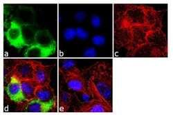
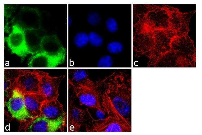
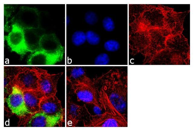
- Immunofluorescence analysis of Cyclin D1 was done on 70% confluent log phase SH-SY5Y cells. The cells were fixed with 4% paraformaldehyde for 10 minutes, permeabilized with 0.1% Triton™ X-100 for 10 minutes, and blocked with 1% BSA for 1 hour at room temperature. The cells were labeled with Cyclin D1 (DCS-6) Mouse Monoclonal Antibody (Product # AHF0082) at 2 µg/mL in 0.1% BSA and incubated for 3 hours at room temperature and then labeled with Goat anti-Mouse IgG (H+L) Superclonal™ Secondary Antibody, Alexa Fluor® 488 conjugate (Product # A28175) at a dilution of 1:2000 for 45 minutes at room temperature (Panel a: green). Nuclei (Panel b: blue) were stained with SlowFade® Gold Antifade Mountant with DAPI (Product # S36938). F-actin (Panel c: red) was stained with Alexa Fluor® 555 Rhodamine Phalloidin (Product # R415, 1:300). Panel d is a merged image showing cytoplasmic localization. Panel e is a no primary antibody control. The images were captured at 60X magnification.
- Submitted by
- Invitrogen Antibodies (provider)
- Main image

- Experimental details
- Immunofluorescence analysis of Cyclin D1 was done on 70% confluent log phase SH-SY5Y cells. The cells were fixed with 4% paraformaldehyde for 10 minutes, permeabilized with 0.1% Triton™ X-100 for 10 minutes, and blocked with 1% BSA for 1 hour at room temperature. The cells were labeled with Cyclin D1 (DCS-6) Mouse Monoclonal Antibody (Product # AHF0082) at 2 µg/mL in 0.1% BSA and incubated for 3 hours at room temperature and then labeled with Goat anti-Mouse IgG (H+L) Superclonal™ Secondary Antibody, Alexa Fluor® 488 conjugate (Product # A28175) at a dilution of 1:2000 for 45 minutes at room temperature (Panel a: green). Nuclei (Panel b: blue) were stained with SlowFade® Gold Antifade Mountant with DAPI (Product # S36938). F-actin (Panel c: red) was stained with Alexa Fluor® 555 Rhodamine Phalloidin (Product # R415, 1:300). Panel d is a merged image showing cytoplasmic localization. Panel e is a no primary antibody control. The images were captured at 60X magnification.
Supportive validation
- Submitted by
- Invitrogen Antibodies (provider)
- Main image

- Experimental details
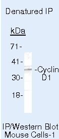
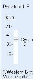
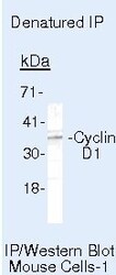
- Immunoprecipitation of Cyclin D1 in denatured mouse MAD109 cells using a Cyclin D1 monoclonal antibody (Product # AHF0082).
- Submitted by
- Invitrogen Antibodies (provider)
- Main image
- Experimental details
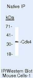
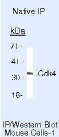
- Immunoprecipitation of Cyclin D1 in native mouse MAD109 cells using a Cyclin D1 monoclonal antibody (Product # AHF0082).

Supportive validation
- Submitted by
- Invitrogen Antibodies (provider)
- Main image
- Experimental details
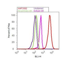
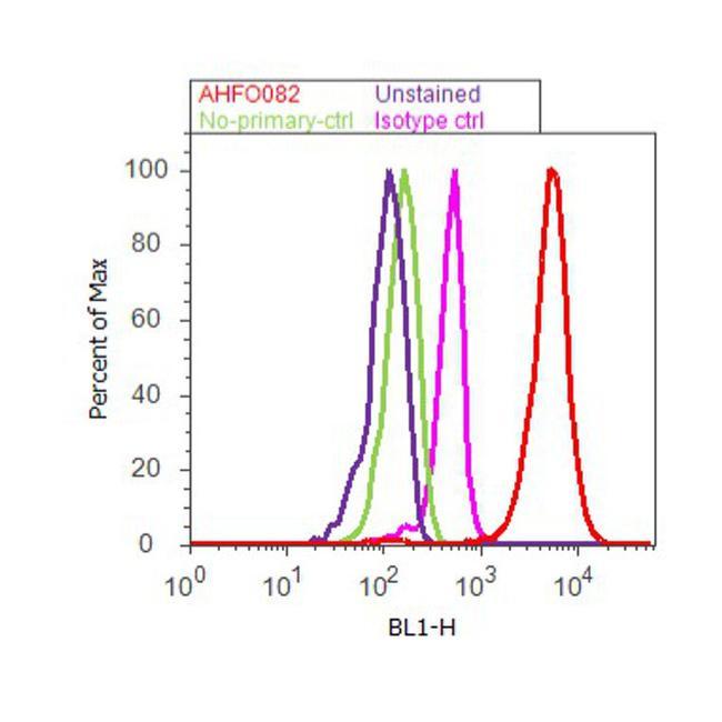
- Flow cytometry analysis of CYCLIND1 was done on SH-SY5Y cells. Cells were fixed with 70% ethanol for 10 minutes, permeabilized with 0.25% Triton™ X-100 for 20 minutes, and blocked with 5% BSA for 30 minutes at room temperature. Cells were labeled with CYCLIND1 Mouse Monoclonal Antibody (AHF0082, red histogram) or with mouse isotype control (pink histogram) at 3-5 ug/million cells in 2.5% BSA. After incubation at room temperature for 2 hours, the cells were labeled with Alexa Fluor® 488 Rabbit Anti-Mouse Secondary Antibody (A11059) at a dilution of 1:400 for 30 minutes at room temperature. The representative 10,000 cells were acquired and analyzed for each sample using an Attune® Acoustic Focusing Cytometer. The purple histogram represents unstained control cells and the green histogram represents no-primary-antibody control.
Supportive validation
- Submitted by
- Invitrogen Antibodies (provider)
- Main image

- Experimental details
- Immunoprecipitation of Cyclin D1 in denatured mouse MAD109 cells using a Cyclin D1 monoclonal antibody (Product # AHF0082).
- Submitted by
- Invitrogen Antibodies (provider)
- Main image
- Experimental details
- NULL
- Submitted by
- Invitrogen Antibodies (provider)
- Main image
- Experimental details
- Immunoprecipitation of Cyclin D1 in denatured mouse MAD109 cells using a Cyclin D1 monoclonal antibody (Product # AHF0082).

- Submitted by
- Invitrogen Antibodies (provider)
- Main image

- Experimental details
- Immunoprecipitation of Cyclin D1 in native mouse MAD109 cells using a Cyclin D1 monoclonal antibody (Product # AHF0082).
- Submitted by
- Invitrogen Antibodies (provider)
- Main image

- Experimental details
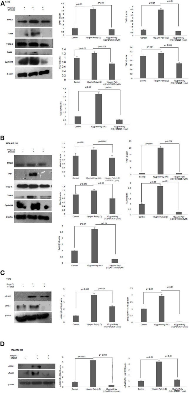
- Figure 5 Western blotting for the expression of signaling protein. (A,B) Cell lysate were collected and subjected to Western blot assay to estimate the level of expression of interleukin 1 receptor-associated kinase 1 (IRAK1), transforming growth factor beta-activated kinase 1 (TAK1), TGF-beta-activated kinase 1 (TAB1), TNF receptor-associated factor 6 (TRAF6), and cyclin D1. (C,D) Expression of pIRAK1 and pTAK1. beta-actin was used as loading control. The respective bar graphs are presented as densitometry analysis as mean +- SD of experiments ( p < 0.05 is treated as significant).