Explore
 Validate
 Learn
Learn Western blot
 ELISA
ELISAAntibody data
- Antibody Data
- Antigen structure
- References [0]
- Comments [0]
- Validations
- Western blot [2]
- Immunocytochemistry [3]
- Immunohistochemistry [5]
- Flow cytometry [1]
Submit
Validation data
Reference
Comment
Report error
- Product number
- BS-0623R - Provider product page

- Provider
- Invitrogen Antibodies
- Product name
- Cyclin D1 Polyclonal Antibody
- Antibody type
- Polyclonal
- Antigen
- Synthetic peptide
- Reactivity
- Human, Mouse, Rat, Bovine, Rabbit
- Host
- Rabbit
- Isotype
- IgG
- Vial size
- 100 µL
- Concentration
- 1 mg/mL
- Storage
- -20° C, Avoid Freeze/Thaw Cycles
No comments: Submit comment
Supportive validation
- Submitted by
- Invitrogen Antibodies (provider)
- Main image
- Experimental details
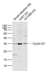
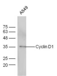
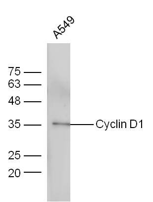
- A549 cells probed with Anti-Cyclin D1 Polyclonal Antibody (bs-0623R) at 1:300 overnight in 4°C. Followed by conjugation to the secondary antibody (bs-0295G-HRP) at 1:5000 90 min in 37°C.
- Submitted by
- Invitrogen Antibodies (provider)
- Main image
- Experimental details
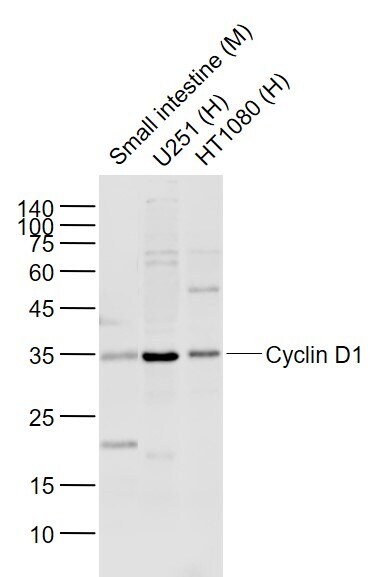
- A549 cells probed with Anti-Cyclin D1 Polyclonal Antibody (bs-0623R) at 1:300 overnight in 4°C. Followed by conjugation to the secondary antibody (bs-0295G-HRP) at 1:5000 90 min in 37°C.
Supportive validation
- Submitted by
- Invitrogen Antibodies (provider)
- Main image
- Experimental details
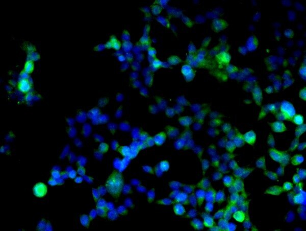
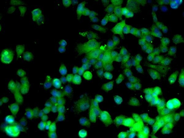
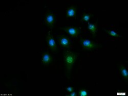
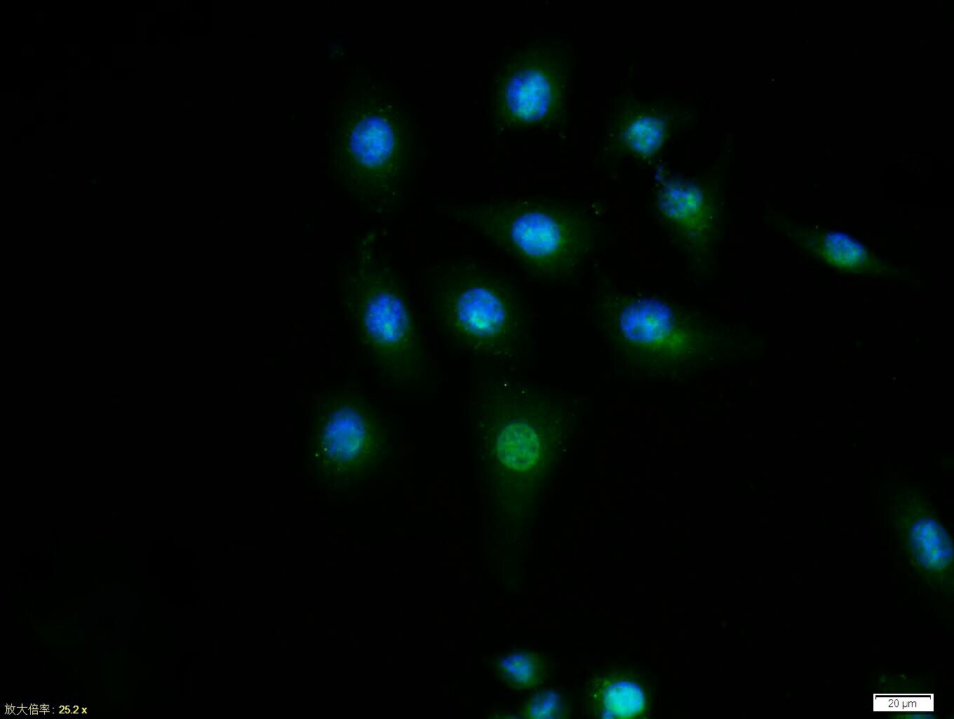
- Image provided by One World Lab validation program. HEK293 cells probed with Rabbit Anti-Cyclin D1 Polyclonal Antibody (bs-0623R) at 1:50 for 60 minutes at room temperature followed by Goat Anti-Rabbit IgG (H+L) Alexa Fluor 488 Conjugated secondary antibody.
- Submitted by
- Invitrogen Antibodies (provider)
- Main image
- Experimental details
- Image provided by One World Lab validation program. HEK293 cells probed with Rabbit Anti-Cyclin D1 Polyclonal Antibody (bs-0623R) at 1:50 for 60 minutes at room temperature followed by Goat Anti-Rabbit IgG (H+L) Alexa Fluor 488 Conjugated secondary antibody.
- Submitted by
- Invitrogen Antibodies (provider)
- Main image
- Experimental details
- Image provided by One World Lab validation program. HEK293 cells probed with Rabbit Anti-Cyclin D1 Polyclonal Antibody (bs-0623R) at 1:50 for 60 minutes at room temperature followed by Goat Anti-Rabbit IgG (H+L) Alexa Fluor 488 Conjugated secondary antibody.
Supportive validation
- Submitted by
- Invitrogen Antibodies (provider)
- Main image
- Experimental details
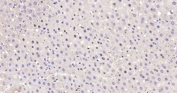
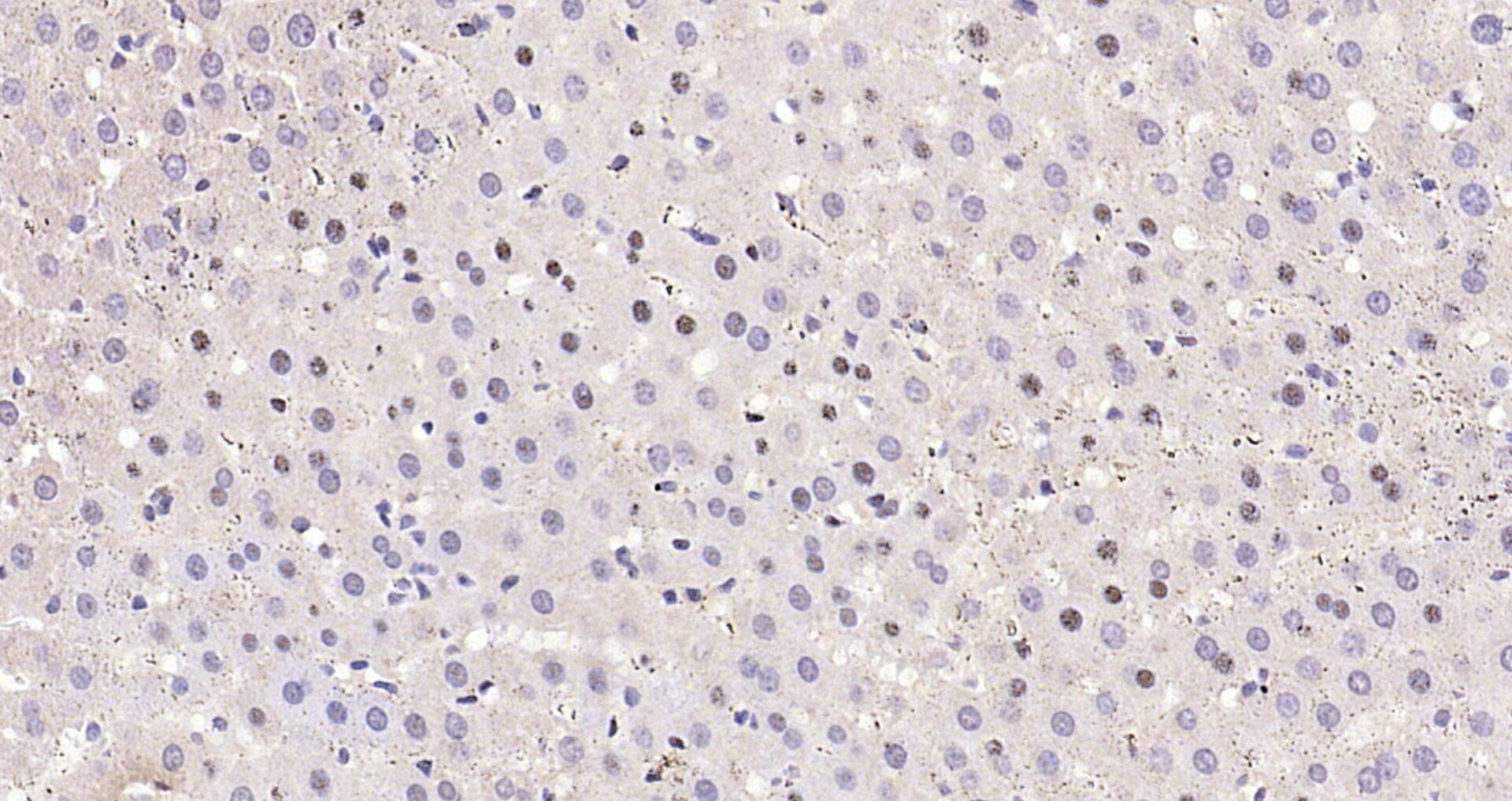
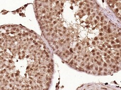
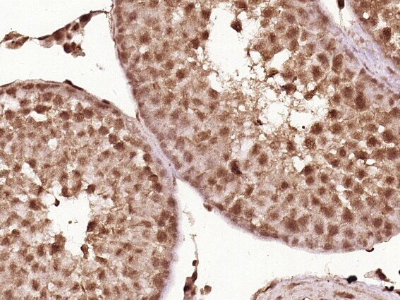
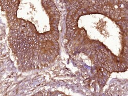
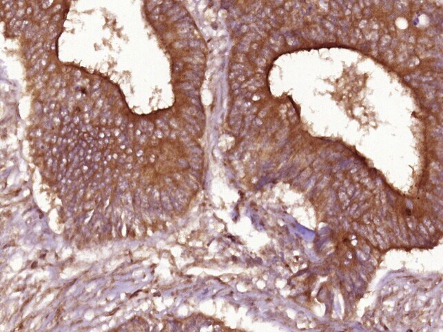
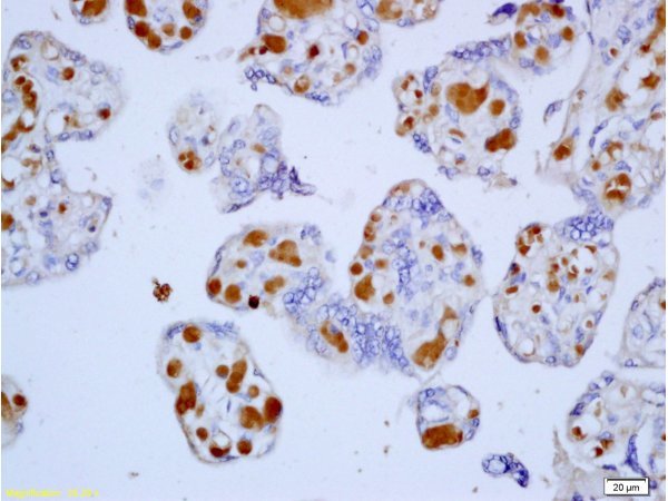
- Formalin-fixed and paraffin embedded human placenta tissue labeled with Anti-Cyclin D1 Polyclonal Antibody, Unconjugated (bs-0623R) at 1:200, followed by conjugation to the secondary antibody and DAB staining.
- Submitted by
- Invitrogen Antibodies (provider)
- Main image
- Experimental details
- Formalin-fixed and paraffin embedded human placenta tissue labeled with Anti-Cyclin D1 Polyclonal Antibody, Unconjugated (bs-0623R) at 1:200, followed by conjugation to the secondary antibody and DAB staining.
- Submitted by
- Invitrogen Antibodies (provider)
- Main image
- Experimental details
- Formalin-fixed and paraffin embedded human placenta tissue labeled with Anti-Cyclin D1 Polyclonal Antibody, Unconjugated (bs-0623R) at 1:200, followed by conjugation to the secondary antibody and DAB staining.
- Submitted by
- Invitrogen Antibodies (provider)
- Main image
- Experimental details
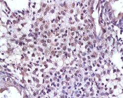
- Formalin-fixed and paraffin embedded human placenta tissue labeled with Anti-Cyclin D1 Polyclonal Antibody, Unconjugated (bs-0623R) at 1:200, followed by conjugation to the secondary antibody and DAB staining.
- Submitted by
- Invitrogen Antibodies (provider)
- Main image
- Experimental details
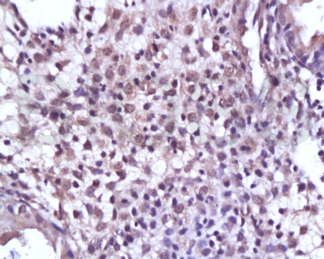
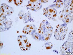
- Formalin-fixed and paraffin embedded human placenta tissue labeled with Anti-Cyclin D1 Polyclonal Antibody, Unconjugated (bs-0623R) at 1:200, followed by conjugation to the secondary antibody and DAB staining.
Supportive validation
- Submitted by
- Invitrogen Antibodies (provider)
- Main image
- Experimental details
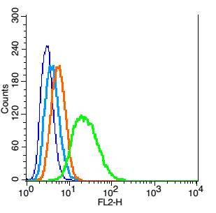
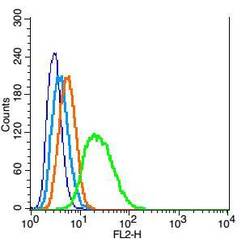
- RSC96 cells probed with Cyclin D1 Polyclonal Antibody, Unconjugated (bs-0623R) at 1:100 for 30 minutes followed by incubation with a conjugated secondary (PE Conjugated) (green) for 30 minutes compared to control cells (blue), secondary only (light blue) and isotype control (orange).