Explore
 Validate
 Learn
Learn Western blot
Antibody data
- Antibody Data
- Antigen structure
- References [9]
- Comments [0]
- Validations
- Western blot [2]
- Immunohistochemistry [3]
Submit
Validation data
Reference
Comment
Report error
- Product number
- PA5-16777 - Provider product page

- Provider
- Invitrogen Antibodies
- Product name
- Cyclin D1 Polyclonal Antibody
- Antibody type
- Polyclonal
- Antigen
- Synthetic peptide
- Description
- PA5-16777 targets Cyclin D1/Bcl-1 in IHC (P), IP, and WB applications and shows reactivity with Human, mouse, and Rat samples.
- Concentration
- 0.2 mg/mL
Submitted references Regulation of PTEN translation by PI3K signaling maintains pathway homeostasis.
Downregulation of ROS1 enhances the therapeutic efficacy of arsenic trioxide in acute myeloid leukemia cell lines.
Aberrant Activation of the RANK Signaling Receptor Induces Murine Salivary Gland Tumors.
Chronic Deletion and Acute Knockdown of Parkin Have Differential Responses to Acetaminophen-induced Mitophagy and Liver Injury in Mice.
Synergistic signaling of KRAS and thyroid hormone receptor β mutants promotes undifferentiated thyroid cancer through MYC up-regulation.
Progesterone receptor activation downregulates GATA3 by transcriptional repression and increased protein turnover promoting breast tumor growth.
Smoothened controls cyclin D2 expression and regulates the generation of intermediate progenitors in the developing cortex.
Antitumor effect of metformin in esophageal cancer: in vitro study.
Epithelial progesterone receptor exhibits pleiotropic roles in uterine development and function.
Mukherjee R, Vanaja KG, Boyer JA, Gadal S, Solomon H, Chandarlapaty S, Levchenko A, Rosen N
Molecular cell 2021 Feb 18;81(4):708-723.e5
Molecular cell 2021 Feb 18;81(4):708-723.e5
Downregulation of ROS1 enhances the therapeutic efficacy of arsenic trioxide in acute myeloid leukemia cell lines.
Li J
Oncology letters 2018 Jun;15(6):9392-9396
Oncology letters 2018 Jun;15(6):9392-9396
Aberrant Activation of the RANK Signaling Receptor Induces Murine Salivary Gland Tumors.
Szwarc MM, Kommagani R, Jacob AP, Dougall WC, Ittmann MM, Lydon JP
PloS one 2015;10(6):e0128467
PloS one 2015;10(6):e0128467
Chronic Deletion and Acute Knockdown of Parkin Have Differential Responses to Acetaminophen-induced Mitophagy and Liver Injury in Mice.
Williams JA, Ni HM, Haynes A, Manley S, Li Y, Jaeschke H, Ding WX
The Journal of biological chemistry 2015 Apr 24;290(17):10934-46
The Journal of biological chemistry 2015 Apr 24;290(17):10934-46
Synergistic signaling of KRAS and thyroid hormone receptor β mutants promotes undifferentiated thyroid cancer through MYC up-regulation.
Zhu X, Zhao L, Park JW, Willingham MC, Cheng SY
Neoplasia (New York, N.Y.) 2014 Sep;16(9):757-69
Neoplasia (New York, N.Y.) 2014 Sep;16(9):757-69
Progesterone receptor activation downregulates GATA3 by transcriptional repression and increased protein turnover promoting breast tumor growth.
Izzo F, Mercogliano F, Venturutti L, Tkach M, Inurrigarro G, Schillaci R, Cerchietti L, Elizalde PV, Proietti CJ
Breast cancer research : BCR 2014 Dec 6;16(6):491
Breast cancer research : BCR 2014 Dec 6;16(6):491
Smoothened controls cyclin D2 expression and regulates the generation of intermediate progenitors in the developing cortex.
Komada M, Iguchi T, Takeda T, Ishibashi M, Sato M
Neuroscience letters 2013 Jun 28;547:87-91
Neuroscience letters 2013 Jun 28;547:87-91
Antitumor effect of metformin in esophageal cancer: in vitro study.
Kobayashi M, Kato K, Iwama H, Fujihara S, Nishiyama N, Mimura S, Toyota Y, Nomura T, Nomura K, Tani J, Miyoshi H, Kobara H, Mori H, Murao K, Masaki T
International journal of oncology 2013 Feb;42(2):517-24
International journal of oncology 2013 Feb;42(2):517-24
Epithelial progesterone receptor exhibits pleiotropic roles in uterine development and function.
Franco HL, Rubel CA, Large MJ, Wetendorf M, Fernandez-Valdivia R, Jeong JW, Spencer TE, Behringer RR, Lydon JP, Demayo FJ
FASEB journal : official publication of the Federation of American Societies for Experimental Biology 2012 Mar;26(3):1218-27
FASEB journal : official publication of the Federation of American Societies for Experimental Biology 2012 Mar;26(3):1218-27
No comments: Submit comment
Supportive validation
- Submitted by
- Invitrogen Antibodies (provider)
- Main image
- Experimental details
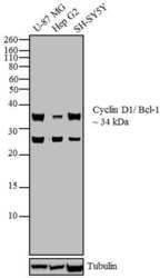
- Western blot analysis was performed using whole cell extracts (30 µg) of U-87 MG (Lane 1), Hep G2 (Lane 2) and SH-SY5Y (Lane 3). The blots was probed with Anti-Cyclin D1/ Bcl-1 Rabbit Polyclonal Antibody (Product # PA5-16777, 2 µg/mL) and detected by chemiluminescence using Goat anti-Rabbit IgG (H+L) Superclonal™ Secondary Antibody, HRP conjugate (Product # A27036, 0.4µg/mL, 1:2500 dilution). A ~ 34 kDa band corresponding to Cyclin D1/ Bcl-1 was observed across cell lines tested. Known quantity of protein samples were electrophoresed using Novex® NuPAGE®12 % Bis-Tris gel (Product # NP0342BOX), XCell SureLock™ Electrophoresis System (Product # EI0002) and Novex® Sharp Pre-Stained Protein Standard (Product # LC5800). Resolved proteins were then transferred onto a nitrocellulose membrane with iBlot® 2 Dry Blotting System (Product # IB21001). The membrane was probed with the relevant primary and secondary Antibody using iBind™ Flex Western Starter Kit (Product # SLF2000S). Chemiluminescent detection was performed using Pierce™ ECL Western Blotting Substrate (Product # 32106).

- Submitted by
- Invitrogen Antibodies (provider)
- Main image

- Experimental details
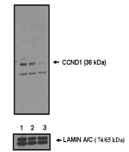
- Western blot analysis of CCND1 was performed with 10 µg of A549 cells transfected with Transfection Reagent alone (Lane 1), 100nM Non-Targeting control siRNA (Lane 2), or 100nM siRNA against CCND1 (Lane 3). Proteins were resolved using a NuPAGE® Novex 4-12% Bis-Tris Gel (Product # NP0322BOX), XCell SureLock™ Electrophoresis System (Product # EI0002), and a protein size ladder. Proteins were wet transferred to a Pierce Nitrocellulose Membrane (Product # 88025) OR Pierce PVDF Membrane (Product # 88518) and blocked with Pierce Starting Block T20 (PBS) Blocking Buffer (Product # 37539) for 1 hour at room temperature. CCND1 was detected at ~ 36 kDa using CCND1 Rabbit polyclonal antibody (Product # PA5-16777) diluted in Pierce Starting Block T20 (PBS) Blocking Buffer 4°C overnight on a rocking platform. Pierce Goat Anti-Rabbit (Product # 31461) HRP-Conjugated Antibodies at a 1:2500 dilution were used and chemiluminescent detection was performed using Pierce Supersignal West Dura Maximum Sensitivity Substrate (Product # 37071). Relative density of the bands normalized to Lamin A/C (74/65 kDa). CCND1 Antibody (Product # PA5-16777) confirms silencing of CCND1 expression.
Supportive validation
- Submitted by
- Invitrogen Antibodies (provider)
- Main image

- Experimental details
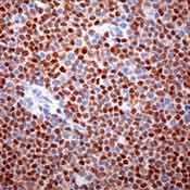
- Formalin-fixed, paraffin-embedded human mantle cell lymphoma stained with Cyclin D1 antibody using peroxidase-conjugate and DAB. Note nuclear staining of tumor cells
- Submitted by
- Invitrogen Antibodies (provider)
- Main image
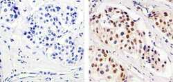
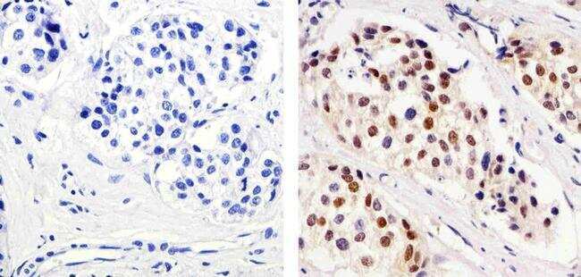
- Experimental details
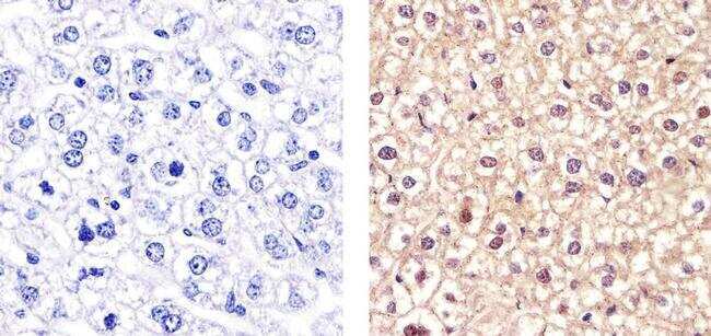
- Immunohistochemistry analysis of Cyclin D1/Bcl-1 showing staining in the nucleus of paraffin-embedded human breast carcinoma (right) compared to a negative control without primary antibody (left). To expose target proteins, antigen retrieval was performed using 10mM sodium citrate (pH 6.0), microwaved for 8-15 min. Following antigen retrieval, tissues were blocked in 3% H2O2-methanol for 15 min at room temperature, washed with ddH2O and PBS, and then probed with a Cyclin D1/Bcl-1 Rabbit Polyclonal Antibody (Product # PA5-16777) diluted in 3% BSA-PBS at a dilution of 1:100 for 1 hour at 37°C in a humidified chamber. Tissues were washed extensively in PBST and detection was performed using an HRP-conjugated secondary antibody followed by colorimetric detection using a DAB kit. Tissues were counterstained with hematoxylin and dehydrated with ethanol and xylene to prep for mounting.
- Submitted by
- Invitrogen Antibodies (provider)
- Main image

- Experimental details
- Immunohistochemistry analysis of Cyclin D1/Bcl-1 showing staining in the nucleus and weak cytoplasm of paraffin-embedded mouse liver tissue (right) compared to a negative control without primary antibody (left). To expose target proteins, antigen retrieval was performed using 10mM sodium citrate (pH 6.0), microwaved for 8-15 min. Following antigen retrieval, tissues were blocked in 3% H2O2-methanol for 15 min at room temperature, washed with ddH2O and PBS, and then probed with a Cyclin D1/Bcl-1 Rabbit Polyclonal Antibody (Product # PA5-16777) diluted in 3% BSA-PBS at a dilution of 1:20 for 1 hour at 37°C in a humidified chamber. Tissues were washed extensively in PBST and detection was performed using an HRP-conjugated secondary antibody followed by colorimetric detection using a DAB kit. Tissues were counterstained with hematoxylin and dehydrated with ethanol and xylene to prep for mounting.