 Explore
 Validate
 Learn
Learn Western blot
Antibody data
- Antibody Data
- Antigen structure
- References [4]
- Comments [0]
- Validations
- Western blot [7]
- Immunocytochemistry [1]
- Immunoprecipitation [1]
- Immunohistochemistry [2]
- Chromatin Immunoprecipitation [1]
Submit
Validation data
Reference
Comment
Report error
- Product number
- GTX100531 - Provider product page

- Provider
- GeneTex
- Proper citation
- GeneTex Cat#GTX100531, RRID:AB_10624270
- Product name
- MDM2 antibody
- Antibody type
- Polyclonal
- Reactivity
- Human, Mouse
- Host
- Rabbit
Submitted references Mechanisms of Targeting the MDM2-p53-FOXM1 Axis in Well-Differentiated Intestinal Neuroendocrine Tumors.
Transketolase Regulates the Metabolic Switch to Control Breast Cancer Cell Metastasis via the α-Ketoglutarate Signaling Pathway.
Autoantibody to MDM2: A Potential Serological Marker of Systemic Lupus Erythematosus.
Autoantibodies response to MDM2 and p53 in the immunodiagnosis of esophageal squamous cell carcinoma.
Briest F, Grass I, Sedding D, Möbs M, Christen F, Benecke J, Fuchs K, Mende S, Kaemmerer D, Sänger J, Kunze A, Geisler C, Freitag H, Lewens F, Worpenberg L, Iwaszkiewicz S, Siegmund B, Walther W, Hummel M, Grabowski P
Neuroendocrinology 2018;107(1):1-23
Neuroendocrinology 2018;107(1):1-23
Transketolase Regulates the Metabolic Switch to Control Breast Cancer Cell Metastasis via the α-Ketoglutarate Signaling Pathway.
Tseng CW, Kuo WH, Chan SH, Chan HL, Chang KJ, Wang LH
Cancer research 2018 Jun 1;78(11):2799-2812
Cancer research 2018 Jun 1;78(11):2799-2812
Autoantibody to MDM2: A Potential Serological Marker of Systemic Lupus Erythematosus.
Liu Y, Dai L, Liu W, Shi G, Zhang J
Journal of immunology research 2015;2015:963568
Journal of immunology research 2015;2015:963568
Autoantibodies response to MDM2 and p53 in the immunodiagnosis of esophageal squamous cell carcinoma.
Chai Y, Peng B, Dai L, Qian W, Zhang Y, Zhang JY
Scandinavian journal of immunology 2014 Nov;80(5):362-8
Scandinavian journal of immunology 2014 Nov;80(5):362-8
No comments: Submit comment
Supportive validation
- Submitted by
- GeneTex (provider)
- Main image
- Experimental details
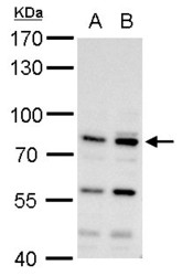
- MDM2 antibody detects MDM2 protein by western blot analysis.A. 30 ?g H1299 whole cell lysate/extract B. 30 ?g HCT116 whole cell lysate/extract7.5% SDS-PAGEMDM2 antibody (GTX100531) dilution: 1:500 The HRP-conjugated anti-rabbit IgG antibody (GTX213110-01) was used to detect the primary antibody.

- Submitted by
- GeneTex (provider)
- Main image
- Experimental details
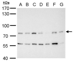
- MDM2 antibody detects MDM2 protein by western blot analysis.A. 30 ?g Neuro2A whole cell lysate/extract B. 30 ?g GL261 whole cell lysate/extract C. 30 ?g C8D30 whole cell lysate/extract D. 30 ?g NIH-3T3 whole cell lysate/extract E. 30 ?g BCL-1 whole cell lysate/extract F. 30 ?g Raw264.7 whole cell lysate/extract G. 30 ?g C2C12 whole cell lysate/extract7.5% SDS-PAGEMDM2 antibody (GTX100531) dilution: 1:1000 The HRP-conjugated anti-rabbit IgG antibody (GTX213110-01) was used to detect the primary antibody.

- Submitted by
- GeneTex (provider)
- Main image
- Experimental details
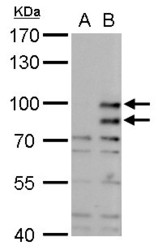
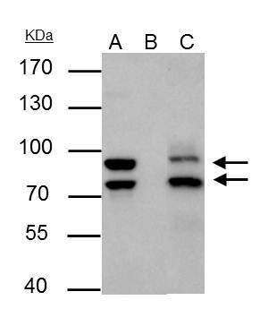
- MDM2 antibody detects MDM2 protein by western blot analysis.A. 30 ?g 293T whole cell lysate/extract B. 30 ?g whole cell lysate/extract of mouse MDM2-transfected 293T cells7.5% SDS-PAGEMDM2 antibody (GTX100531) dilution: 1:1000 The HRP-conjugated anti-rabbit IgG antibody (GTX213110-01) was used to detect the primary antibody.
- Submitted by
- GeneTex (provider)
- Main image

- Experimental details
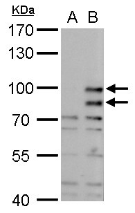
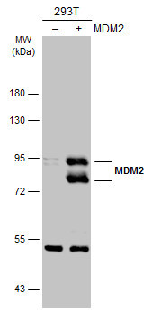
- Non-transfected (¡V) and transfected (+) 293T whole cell extracts (30 ?g) were separated by 7.5% SDS-PAGE, and the membrane was blotted with MDM2 antibody (GTX100531) diluted at 1:2000. The HRP-conjugated anti-rabbit IgG antibody (GTX213110-01) was used to detect the primary antibody.
- Submitted by
- GeneTex (provider)
- Main image
- Experimental details
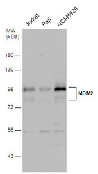
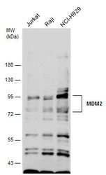
- Various whole cell extracts (30 ?g) were separated by 7.5% SDS-PAGE, and the membrane was blotted with MDM2 antibody (GTX100531) diluted at 1:1000.
- Submitted by
- GeneTex (provider)
- Main image
- Experimental details
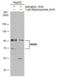
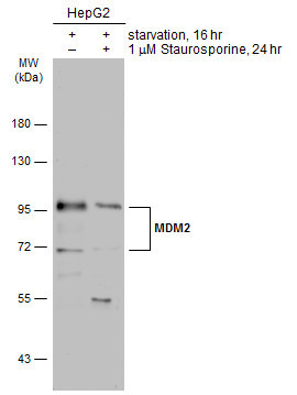
- Untreated (¡V) and treated (+) HepG2 whole cell extracts (30 ?g) were separated by 7.5% SDS-PAGE, and the membrane was blotted with MDM2 antibody (GTX100531) diluted at 1:3000. The HRP-conjugated anti-rabbit IgG antibody (GTX213110-01) was used to detect the primary antibody.
- Submitted by
- GeneTex (provider)
- Main image
- Experimental details
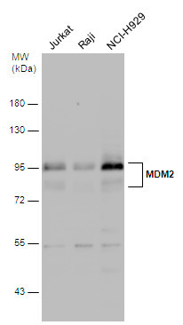
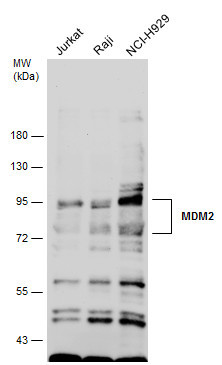
- Various whole cell extracts (30 ?g) were separated by 7.5% SDS-PAGE, and the membrane was blotted with MDM2 antibody (GTX100531) diluted at 1:1000.
Supportive validation
- Submitted by
- GeneTex (provider)
- Main image
- Experimental details
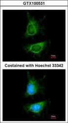
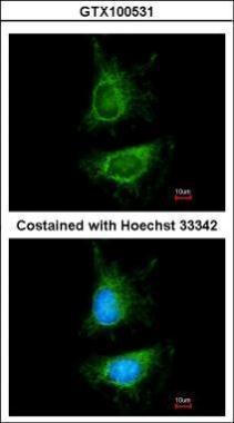
- Immunofluorescence analysis of paraformaldehyde-fixed HeLa, using MDM2(GTX100531) antibody at 1:200 dilution.
Supportive validation
- Submitted by
- GeneTex (provider)
- Main image

- Experimental details
- MDM2 antibody immunoprecipitates MDM2 protein in IP experiments.IP samples: Jurkat whole cell extractA. 30 £gg Jurkat cell whole cell extractB. Control with 4 £gg of preimmune Rabbit IgGC. Immunoprecipitation of MDM2 protein by 4 £gg MDM2 antibody (GTX100531)7.5 % SDS-PAGEThe immunoprecipitated MDM2 protein was detected by MDM2 antibody (GTX100531) diluted at 1:500.[EasyBlot anti-rabbit IgG (GTX221666-01) was used as a secondary reagent]
Supportive validation
- Submitted by
- GeneTex (provider)
- Main image
- Experimental details
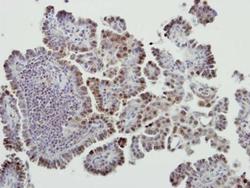
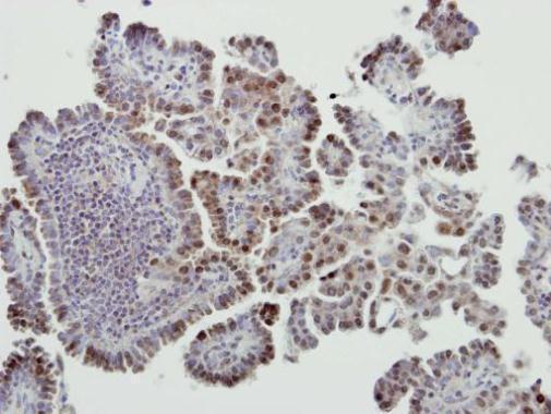
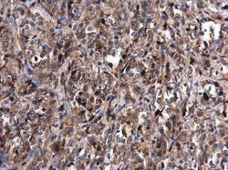
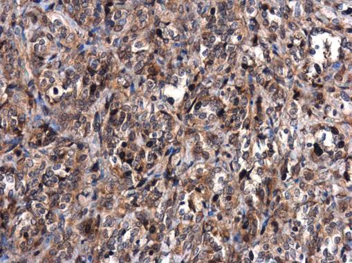
- Immunohistochemical analysis of paraffin-embedded human lung cancer, using MDM2(GTX100531) antibody at 1:500 dilution.
- Submitted by
- GeneTex (provider)
- Main image
- Experimental details
- MDM2 antibody detects MDM2 protein at cytoplasm in human lung by immunohistochemical analysis. Sample: Paraffin-embedded human lung. MDM2 antibody (GTX100531) diluted at 1:500.
Supportive validation
- Submitted by
- GeneTex (provider)
- Main image
- Experimental details
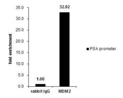
- Cross-linked ChIP was performed with PC-3 chromatin extract and 5 £gg of either control rabbit IgG or anti-MDM2 antibody. The precipitated DNA was detected by PCR with primer set targeting to PSA promoter.
