 Explore
 Validate
 Learn
Learn Western blot
Antibody data
- Antibody Data
- Antigen structure
- References [3]
- Comments [0]
- Validations
- Western blot [1]
Submit
Validation data
Reference
Comment
Report error
- Product number
- PAB10274 - Provider product page

- Provider
- Abnova Corporation
- Proper citation
- Abnova Corporation Cat#PAB10274, RRID:AB_1676440
- Product name
- Mdm2 polyclonal antibody
- Antibody type
- Polyclonal
- Description
- Rabbit polyclonal antibody raised against synthetic peptide of Mdm2.
- Storage
- Store at 4°C. For long term storage store at -20°C.Aliquot to avoid repeated freezing and thawing.
Submitted references Constitutive and DNA damage inducible activation of pig3 and MDM2 genes by tumor-derived p53 mutant C277Y.
High-affinity binding of tumor-suppressor protein p53 and HMGB1 to hemicatenated DNA loops.
Repression of the Arf tumor suppressor by E2F3 is required for normal cell cycle kinetics.
Pospísilová S, Siligan C, Ban J, Jug G, Kovar H
Molecular cancer research : MCR 2004 May;2(5):296-304
Molecular cancer research : MCR 2004 May;2(5):296-304
High-affinity binding of tumor-suppressor protein p53 and HMGB1 to hemicatenated DNA loops.
Stros M, Muselíková-Polanská E, Pospísilová S, Strauss F
Biochemistry 2004 Jun 8;43(22):7215-25
Biochemistry 2004 Jun 8;43(22):7215-25
Repression of the Arf tumor suppressor by E2F3 is required for normal cell cycle kinetics.
Aslanian A, Iaquinta PJ, Verona R, Lees JA
Genes & development 2004 Jun 15;18(12):1413-22
Genes & development 2004 Jun 15;18(12):1413-22
No comments: Submit comment
Supportive validation
- Submitted by
- Abnova Corporation (provider)
- Main image
- Experimental details
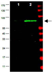
- Western blot using Mdm2 polyclonal antibody (Cat # PAB10274) is shown to detect a band (arrow) corresponding to mouse Mdm2 protein present in mouse MEF cells (Lane 2), but not human kidney HEK293 cells (lane1).Approximately 35 ug of lysate was separated by 4-20% Tris Glycine SDS-PAGE.After blocking the membrane with 5% normal goat serum, 0.5% BLOTTO in PBS, the membrane was probed for overnight at 4° with the primary antibody diluted to 1:500 in 1% normal goat serum, 0.1% BLOTTO in PBS.The membrane was washed and reacted with a 1 : 10,000 dilution of IRDye™800 conjugated Gt-a-Rabbit IgG [H&L] for 45 min at roomtemperature (800 nm channel, green).Molecular weight estimation was made by comparison to prestained MW markers indicated at the right (700 nm channel, red).IRDye™800 fluorescence image was captured using the Odyssey® Infrared Imaging System developed by LI-COR.IRDye is a trademark of LI-COR, Inc.
