Explore
 Validate
 Learn
LearnPA5-97565
antibody from Invitrogen Antibodies
Targeting: CD59
16.3A5, EJ16, EJ30, EL32, G344, MIC11, MIN1, MIN2, MIN3, MSK21, p18-20
 Western blot
 ELISA
ELISA Immunocytochemistry
Antibody data
- Antibody Data
- Antigen structure
- References [1]
- Comments [0]
- Validations
- Immunocytochemistry [3]
- Immunohistochemistry [1]
- Other assay [1]
Submit
Validation data
Reference
Comment
Report error
- Product number
- PA5-97565 - Provider product page

- Provider
- Invitrogen Antibodies
- Product name
- CD59 Polyclonal Antibody
- Antibody type
- Polyclonal
- Antigen
- Recombinant full-length protein
- Reactivity
- Human, Mouse
- Host
- Rabbit
- Isotype
- IgG
- Vial size
- 100 μg
- Concentration
- 1 mg/mL
- Storage
- -20°C or -80°C if preferred
Submitted references Proteome Profile of Myelin in the Zebrafish Brain.
Siems SB, Jahn O, Hoodless LJ, Jung RB, Hesse D, Möbius W, Czopka T, Werner HB
Frontiers in cell and developmental biology 2021;9:640169
Frontiers in cell and developmental biology 2021;9:640169
No comments: Submit comment
Supportive validation
- Submitted by
- Invitrogen Antibodies (provider)
- Main image
- Experimental details
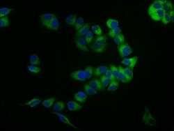
- Immunofluorescent analysis of CD59 in PC-3 cells using a CD59 polyclonal antibody (Product # PA5-97565) at a dilution of 1:133. The cells were fixed in 4% formaldehyde, permeabilized using 0.2% Triton X-100 and blocked in 10% normal Goat Serum. The cells were then incubated with the antibody overnight at 4°C. The secondary antibody was Alexa Fluor 488-congugated Goat Anti-Rabbit IgG(H+L). Cells were counter-stained with DAPI.

- Submitted by
- Invitrogen Antibodies (provider)
- Main image

- Experimental details
- Immunofluorescent analysis of CD59 in PC-3 cells using a CD59 polyclonal antibody (Product # PA5-97565) at a dilution of 1:133. The cells were fixed in 4% formaldehyde, permeabilized using 0.2% Triton X-100 and blocked in 10% normal Goat Serum. The cells were then incubated with the antibody overnight at 4°C. The secondary antibody was Alexa Fluor 488-congugated Goat Anti-Rabbit IgG(H+L). Cells were counter-stained with DAPI.
- Submitted by
- Invitrogen Antibodies (provider)
- Main image
- Experimental details
- Immunofluorescent analysis of CD59 in PC-3 cells using a CD59 polyclonal antibody (Product # PA5-97565) at a dilution of 1:133. The cells were fixed in 4% formaldehyde, permeabilized using 0.2% Triton X-100 and blocked in 10% normal Goat Serum. The cells were then incubated with the antibody overnight at 4°C. The secondary antibody was Alexa Fluor 488-congugated Goat Anti-Rabbit IgG(H+L). Cells were counter-stained with DAPI.

Supportive validation
- Submitted by
- Invitrogen Antibodies (provider)
- Main image
- Experimental details
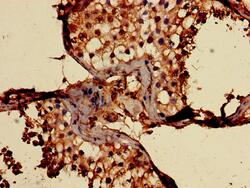
- Immunohistochemical analysis of CD59 in paraffin embedded human testis tissue using a CD59 polyclonal antibody (Product # PA5-97565) at a dilution of 1:100.

Supportive validation
- Submitted by
- Invitrogen Antibodies (provider)
- Main image
- Experimental details
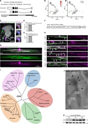
- FIGURE 5 Expression and subcellular localization of CD59 in the zebrafish spinal cord. (A) Gene structure of zebrafish cd59 . Filled boxes indicate the open reading frame. (B) tSNE plot showing cells displaying high cd59 transcript abundance (labeled in red) by scRNAseq (). Cells with low or absent cd59 expression are labeled in gray. (B'') Oligodendrocyte lineage cluster layout for the tSNE plot in (B) with the 19 cells in the mature oligodendrocyte cluster (mOL; green) and the combined OPC clusters (gray). Note the overlap between high cd59 expression and the mature oligodendrocyte cluster. (C) Example micrographs of RNAscope in situ hybridization detecting cd59 transcripts in transverse spinal cord sections of Tg(olig1:nls-mApple x mbp:nlsGFP) zebrafish larvae at 5 dpf (scale bar, 10 mum). Magnified images to the right show a Cd59 + (purple) mbp:nls-EGFP (blue) mature oligodendrocyte (individual channels and merge). Seventy-two transverse sections of four animals were taken and imaged for quantification in (D,E) . No Cd59 + cells were found positive for olig1:nls-mApple (green) representing OPCs. (D) Quantification of the percentage of cd59 RNAscope-positive cells that were mature oligodendrocytes (mOL) according to expression of the mbp:nlsEGFP transgene. n = 27 oligodendrocytes from 72 transverse sections taken along the entire spinal cord (anterior, mid-trunk, posterior) of four individual animals were quantified. (E) Quantification of the percentage of mbp:nlsGFP trans