 Explore
 Validate
 Learn
LearnNB500-400
antibody from Novus Biologicals
Targeting: CD59
16.3A5, EJ16, EJ30, EL32, G344, MIC11, MIN1, MIN2, MIN3, MSK21, p18-20
 Western blot
 Immunoprecipitation
Antibody data
- Antibody Data
- Antigen structure
- References [2]
- Comments [0]
- Validations
- Western blot [1]
- Immunohistochemistry [1]
- Flow cytometry [3]
Submit
Validation data
Reference
Comment
Report error
- Product number
- NB500-400 - Provider product page

- Provider
- Novus Biologicals
- Proper citation
- Novus Cat#NB500-400, RRID:AB_10001608
- Product name
- Mouse Monoclonal CD59 Antibody
- Antibody type
- Monoclonal
- Description
- Protein A purified. The antibody MEM-43/5 reacts with well defined epitope (around L33) on CD59 (Protectin), a 18-20 kDa glycosylphosphatidylinositol (GPI)-anchored glycoprotein expressed on all hematopoietic cells
- Reactivity
- Human, Mouse
- Host
- Mouse
- Isotype
- IgG
- Vial size
- 0.1 mg
- Concentration
- 1.0 mg/ml
- Storage
- Store at 4C. Do not freeze.
Submitted references A hypomorphic PIGA gene mutation causes severe defects in neuron development and susceptibility to complement-mediated toxicity in a human iPSC model.
Excessive activation of the alternative complement pathway in autosomal dominant polycystic kidney disease.
Yuan X, Li Z, Baines AC, Gavriilaki E, Ye Z, Wen Z, Braunstein EM, Biesecker LG, Cheng L, Dong X, Brodsky RA
PloS one 2017;12(4):e0174074
PloS one 2017;12(4):e0174074
Excessive activation of the alternative complement pathway in autosomal dominant polycystic kidney disease.
Su Z, Wang X, Gao X, Liu Y, Pan C, Hu H, Beyer RP, Shi M, Zhou J, Zhang J, Serra AL, Wüthrich RP, Mei C
Journal of internal medicine 2014 Nov;276(5):470-85
Journal of internal medicine 2014 Nov;276(5):470-85
No comments: Submit comment
Supportive validation
- Submitted by
- Novus Biologicals (provider)
- Main image
- Experimental details
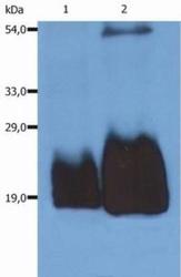
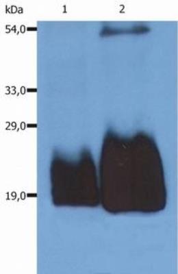
- Western Blot: CD59 Antibody (MEM-43/5) [NB500-400] - Fig. 1. Western Blotting analysis (non-reducing conditions) of whole cell lysate of HPB-ALL human peripheral blood T cell leukemia cell line using anti-CD59 (MEM-43/5). Lane 1: original cell lysate Lane 2: material immunoprecipitated with anti-human CD59 (MEM-43).
Supportive validation
- Submitted by
- Novus Biologicals (provider)
- Main image

- Experimental details
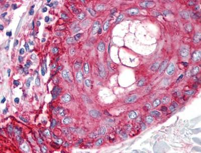
- Immunohistochemistry-Paraffin: CD59 Antibody (MEM-43/5) [NB500-400] - Staining of human skin (paraffin sections) using anti-CD59 (MEM-43/5).
Supportive validation
- Submitted by
- Novus Biologicals (provider)
- Main image
- Experimental details
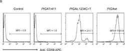
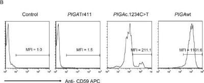
- Flow Cytometry: CD59 Antibody (MEM-43/5) [NB500-400] - Generation of PIGAc.1234C>T mutation using the PiggyBac transposon system. Representative FACS analysis CD59 expression in TF1PIGAnull cells transfected with PB-PIGAwt, PB-PIGAc.1234C>T or PB-PIGAtr411. Transfected TF1PIGAnull cells were stained with an APC-conjugated CD59 antibody to assess PIGA gene expression. Non-transfected TF1PIGAnull cells were used as a control. MFI represents mean fluorescence intensity. Image collected and cropped by CiteAb from the following publication (//dx.plos.org/10.1371/journal.pone.0174074), licensed under a CC-BY licence. Data from the APC-conjugated form of MEM-43/5.
- Submitted by
- Novus Biologicals (provider)
- Main image

- Experimental details
- Flow Cytometry: CD59 Antibody (MEM-43/5) [NB500-400] - The PIGAc.1234C>T mutation increases PIGA function compared to PIGAnull hiPSCs.(A). Representative example of FACS analysis CD59 expression in the three hiPSC lines. Overlay histogram shows that CD59 expression was significantly higher in PIGAc.1234 C>T hiPSCs compared to PIGAnull hiPSCs. MFI was 445.4 in PIGAwt hiPSCs and 332.6 in PIGAc.1234C>T hiPSCs (p>0.05, NS). However, MFI in PIGAc.1234C>T hiPSCs was significantly higher than 17.9 in PIGAnull hiPSCs (*pT hiPSCs. PIGAnull hiPSCs (purple), PIGAc.1234C>T hiPSCs (orange) and PIGAwt hiPSCs (blue). Image collected and cropped by CiteAb from the following publication (//dx.plos.org/10.1371/journal.pone.0174074), licensed under a CC-BY licence. Data from the APC-conjugated form of clone MEM-43/5.

- Submitted by
- Novus Biologicals (provider)
- Main image
- Experimental details
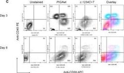
- Flow Cytometry: CD59 Antibody (MEM-43/5) [NB500-400] - The PIGAc.1234C>T mutation does not impair terminal hematopoietic differentiation during mesoderm induction. Representative example of FACS analysis of hematopoietic phenotypes in the EB-BLCs from PIGAwt and PIGAc.1234C>T. The zebra plot shows expression of CD59 (X-axis) and CD45 (Y-axis) after three and eight days of hematopoietic differentiation. Unstained PIGAwt cells were used as a control. Abbreviations: CFU-Macrophage (M); CFU-Granulocyte-Macrophage (GM); committed erythroid BFU-E (BFU) and CFU-E (CFU) progenitors; multipotent progenitor cells CFU-GEMM (GEMM). Image collected and cropped by CiteAb from the following publication (//dx.plos.org/10.1371/journal.pone.0174074), licensed under a CC-BY licence. Data from the APC-conjugated form of anti-CD59 MEM-43/5.
