 Explore
 Validate
 Learn
Learn Western blot
 Immunocytochemistry
Antibody data
- Antibody Data
- Antigen structure
- References [3]
- Comments [0]
- Validations
- Immunocytochemistry [4]
- Immunoprecipitation [1]
- Immunohistochemistry [2]
- Flow cytometry [2]
- Other assay [3]
Submit
Validation data
Reference
Comment
Report error
- Product number
- PA1-109 - Provider product page

- Provider
- Invitrogen Antibodies
- Product name
- PAX5 Polyclonal Antibody
- Antibody type
- Polyclonal
- Antigen
- Purifed from natural sources
- Description
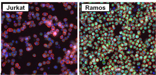
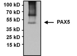
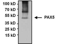
- Western blot analysis of PA1-109 detects a prominent ~45 kDa protein in PAX5 expressing human Ramos, Daudi, Gaji and mouse A20 B-cell lymphoma cells. No PAX5 was detected in negative control Jurkat cells. The 45 kDa band is enriched in immunoprecipitations from Raji lysate, however additional unknown high molecular weight bands are also enriched and detected by PA1-109. PAX5 was also specifically detected by immunofluorescence in the nuclei of Ramos cells compared to negative control Jurkat cells.
- Reactivity
- Human, Mouse
- Host
- Rabbit
- Isotype
- IgG
- Vial size
- 100 μg
- Concentration
- 1 mg/mL
- Storage
- -20°C
Submitted references miR-223-3p Inhibits Human Osteosarcoma Metastasis and Progression by Directly Targeting CDH6.
Cryptotanshinone Protects Cartilage against Developing Osteoarthritis through the miR-106a-5p/GLIS3 Axis.
Relapsed anaplastic lymphoma kinase-positive large B-cell lymphoma expressed cluster of differentiation 4 and cytokeratin: An initially misdiagnosed case corrected by immunoglobulin κ locus gene rearrangement detection.
Ji Q, Xu X, Song Q, Xu Y, Tai Y, Goodman SB, Bi W, Xu M, Jiao S, Maloney WJ, Wang Y
Molecular therapy : the journal of the American Society of Gene Therapy 2018 May 2;26(5):1299-1312
Molecular therapy : the journal of the American Society of Gene Therapy 2018 May 2;26(5):1299-1312
Cryptotanshinone Protects Cartilage against Developing Osteoarthritis through the miR-106a-5p/GLIS3 Axis.
Ji Q, Qi D, Xu X, Xu Y, Goodman SB, Kang L, Song Q, Fan Z, Maloney WJ, Wang Y
Molecular therapy. Nucleic acids 2018 Jun 1;11:170-179
Molecular therapy. Nucleic acids 2018 Jun 1;11:170-179
Relapsed anaplastic lymphoma kinase-positive large B-cell lymphoma expressed cluster of differentiation 4 and cytokeratin: An initially misdiagnosed case corrected by immunoglobulin κ locus gene rearrangement detection.
Shi Y, Li X, Song Y, Zhou L, Feng Q, Wang P, Zhang C, Liu W, Bai Y, Lai Y
Oncology letters 2017 Jul;14(1):787-791
Oncology letters 2017 Jul;14(1):787-791
No comments: Submit comment
Supportive validation
- Submitted by
- Invitrogen Antibodies (provider)
- Main image
- Experimental details
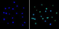
- Immunofluorescent analysis of PAX5 (green) showing staining in the in the nucleus of Ramos cells (right) compared to a negative control without primary antibody (left). Formalin-fixed cells were permeabilized with 0.1% Triton X-100 in TBS for 5-10 minutes and blocked with 3% BSA-PBS for 30 minutes at room temperature. Cells were probed with a PAX5 polyclonal antibody (Product # PA1-109) in 3% BSA-PBS at a dilution of 1:100 and incubated overnight at 4ºC in a humidified chamber. Cells were washed with PBST and incubated with a DyLight-conjugated secondary antibody in PBS at room temperature in the dark.Nuclei (blue) were stained with Hoechst or DAPI. Images were taken at a magnification of 60x.

- Submitted by
- Invitrogen Antibodies (provider)
- Main image

- Experimental details
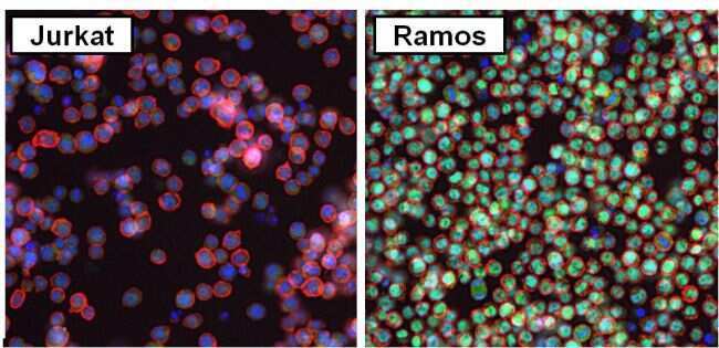
- Immunofluorescent analysis of PAX5 (green) in Ramos and negative control Jurkat cells. Formalin fixed cells were permeabilized with 0.1% Triton X-100 in TBS for 10 minutes at room temperature. Cells were blocked with 2% Blocker BSA (Product # 37525) for 15 minutes at room temperature. Cells were probed with a PAX5 polyclonal antibody (Product # PA1-109) at a dilution of 1:100 for at least 1 hour at room temperature, washed with PBS, and incubated with a DyLight 488-conjugated goat anti-rabbit IgG secondary antibody (Product # 35552) at a dilution of 1:200 for 30 minutes at room temperature. F-Actin (red) was stained with DyLight-554 Phalloidin (Product # 21834) and nuclei (blue) were stained with Hoechst 33342 dye (Product # 62249). Images were taken on a Thermo Scientific ToxInsight Instrument at 20X magnification.
- Submitted by
- Invitrogen Antibodies (provider)
- Main image
- Experimental details
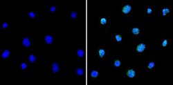
- Immunofluorescent analysis of PAX5 (green) showing staining in the in the nucleus of Raji cells (right) compared to a negative control without primary antibody (left). Formalin-fixed cells were permeabilized with 0.1% Triton X-100 in TBS for 5-10 minutes and blocked with 3% BSA-PBS for 30 minutes at room temperature. Cells were probed with a PAX5 polyclonal antibody (Product # PA1-109) in 3% BSA-PBS at a dilution of 1:100 and incubated overnight at 4ºC in a humidified chamber. Cells were washed with PBST and incubated with a DyLight-conjugated secondary antibody in PBS at room temperature in the dark. Nuclei (blue) were stained with Hoechst or DAPI. Images were taken at a magnification of 60x.

- Submitted by
- Invitrogen Antibodies (provider)
- Main image
- Experimental details
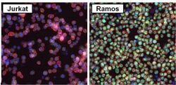
- Immunofluorescent analysis of PAX5 (green) in Ramos and negative control Jurkat cells. Formalin fixed cells were permeabilized with 0.1% Triton X-100 in TBS for 10 minutes at room temperature. Cells were blocked with 2% Blocker BSA (Product # 37525) for 15 minutes at room temperature. Cells were probed with a PAX5 polyclonal antibody (Product # PA1-109) at a dilution of 1:100 for at least 1 hour at room temperature, washed with PBS, and incubated with a DyLight 488-conjugated goat anti-rabbit IgG secondary antibody (Product # 35552) at a dilution of 1:200 for 30 minutes at room temperature. F-Actin (red) was stained with DyLight-554 Phalloidin (Product # 21834) and nuclei (blue) were stained with Hoechst 33342 dye (Product # 62249). Images were taken on a Thermo Scientific ToxInsight Instrument at 20X magnification.
Supportive validation
- Submitted by
- Invitrogen Antibodies (provider)
- Main image
- Experimental details
- Immunoprecipitation of PAX5 was performed using Raji whole cell lysates. Antigen-antibody complexes were formed by incubating 750 µg of lysate with 5 µg of a PAX5 polyclonal antibody (Product # PA1-109) overnight on a rocking platform at 4°C. The immune complexes were captured on 50 µL Protein A/G Agarose (Product # 20421), washed extensively, and eluted with 5X Lane Marker Reducing Sample Buffer (Product # 39000). Samples was resolved on a 4-20% Tris-HCl polyacrylamide gel, transferred to a PVDF membrane, and blocked with 5% BSA/TBS-0.1%Tween-20 for 1 hour. The membrane was probed with a PAX5 polyclonal antibody (Product # PA1-109) at a dilution of 1:1000 overnight rotating at 4°C. Membranes were washed in TBST, and probed with Clean-Blot IP Detection Reagent (Product # 21230) at a dilution of 1:1000 for at least one hour. Chemiluminescent detection was performed using SuperSignal West Pico (Product # 34080).

Supportive validation
- Submitted by
- Invitrogen Antibodies (provider)
- Main image
- Experimental details
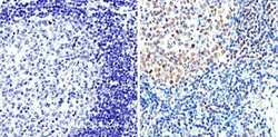
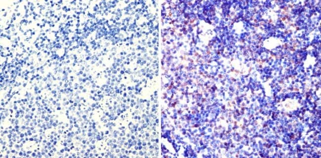
- Immunohistochemistry analysis of PAX5 showing staining in the nucleus of paraffin-embedded human tonsil tissue (right) compared to a negative control without primary antibody (left). To expose target proteins, antigen retrieval was performed using 10mM sodium citrate (pH 6.0), microwaved for 8-15 min. Following antigen retrieval, tissues were blocked in 3% H2O2-methanol for 15 min at room temperature, washed with ddH2O and PBS, and then probed with a PAX5 polyclonal antibody (Product # PA1-109) diluted in 3% BSA-PBS at a dilution of 1:200 overnight at 4°C in a humidified chamber. Tissues were washed extensively in PBST and detection was performed using an HRP-conjugated secondary antibody followed by colorimetric detection using a DAB kit. Tissues were counterstained with hematoxylin and dehydrated with ethanol and xylene to prep for mounting.

- Submitted by
- Invitrogen Antibodies (provider)
- Main image
- Experimental details
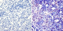
- Immunohistochemistry analysis of PAX5 showing staining in the nucleus of paraffin-embedded mouse lymph node tissue (right) compared to a negative control without primary antibody (left). To expose target proteins, antigen retrieval was performed using 10mM sodium citrate (pH 6.0), microwaved for 8-15 min. Following antigen retrieval, tissues were blocked in 3% H2O2-methanol for 15 min at room temperature, washed with ddH2O and PBS, and then probed with a PAX5 polyclonal antibody (Product # PA1-109) diluted in 3% BSA-PBS at a dilution of 1:100 overnight at 4°C in a humidified chamber. Tissues were washed extensively in PBST and detection was performed using an HRP-conjugated secondary antibody followed by colorimetric detection using a DAB kit. Tissues were counterstained with hematoxylin and dehydrated with ethanol and xylene to prep for mounting.
Supportive validation
- Submitted by
- Invitrogen Antibodies (provider)
- Main image
- Experimental details
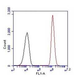
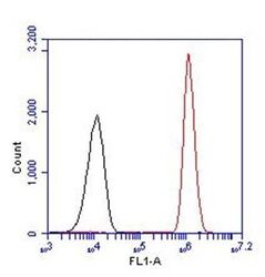
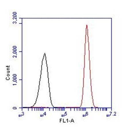
- Flow cytometry analysis of PAX5 (red histogram) on Ramos cells. Cells were harvested, fixed with 4% formaldehyde, permeabilized, washed with PBS, and incubated with a PAX5 polyclonal antibody (Product # PA1-109) at a 1:50 dilution or with PBS alone (black histogram) for 1 hour at room temperature. For flow cytometry analysis, 30-minute incubation with DyLight 488 goat anti-rabbit IgG secondary antibody (Product # 35552) was performed. 30,000 cells were acquired for analysis.
- Submitted by
- Invitrogen Antibodies (provider)
- Main image
- Experimental details
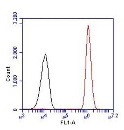
- Flow cytometry analysis of PAX5 (red histogram) on Ramos cells. Cells were harvested, fixed with 4% formaldehyde, permeabilized, washed with PBS, and incubated with a PAX5 polyclonal antibody (Product # PA1-109) at a 1:50 dilution or with PBS alone (black histogram) for 1 hour at room temperature. For flow cytometry analysis, 30-minute incubation with DyLight 488 goat anti-rabbit IgG secondary antibody (Product # 35552) was performed. 30,000 cells were acquired for analysis.
Supportive validation
- Submitted by
- Invitrogen Antibodies (provider)
- Main image
- Experimental details
- Immunoprecipitation of PAX5 was performed using Raji whole cell lysates. Antigen-antibody complexes were formed by incubating 750 µg of lysate with 5 µg of a PAX5 polyclonal antibody (Product # PA1-109) overnight on a rocking platform at 4øC. The immune complexes were captured on 50 µL Protein A/G Agarose (Product # 20421), washed extensively, and eluted with 5X Lane Marker Reducing Sample Buffer (Product # 39000). Samples was resolved on a 4-20% Tris-HCl polyacrylamide gel, transferred to a PVDF membrane, and blocked with 5% BSA/TBS-0.1%Tween-20 for 1 hour. The membrane was probed with a PAX5 polyclonal antibody (Product # PA1-109) at a dilution of 1:1000 overnight rotating at 4øC. Membranes were washed in TBST, and probed with Clean-Blot IP Detection Reagent (Product # 21230) at a dilution of 1:1000 for at least one hour. Chemiluminescent detection was performed using SuperSignal West Pico (Product # 34080).

- Submitted by
- Invitrogen Antibodies (provider)
- Main image
- Experimental details
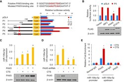
- Figure 4 CTS Promotes the Expression of miR-106a-5p through the Recruitment of PAX5 to the Promoter of miR-106a-5p (A) Luciferase activity of different miR-106a-5p promoter constructs in cultured human chondrocytes with or without CTS stimulation. P1, promoter (-186 bp); P2, promoter (-439 bp); P3, promoter (-761 bp); P4, promoter (-1,000 bp); P5, mutated promoter (-761 bp). (B) Luciferase assay in cultured human chondrocytes infected with FLAG-tagged IRF2 or PAX5 or STAT4 and P5. Immunoblot shows the expression of IRF2, PAX5, and STAT4 with anti-FLAG. (C and D) Cultured human chondrocytes were infected with the PAX5 plasmid (C) and the vector control or shRNA for PAX5 (D), followed by stimulation with CTS as described in the Methods section. Immunoblot indicates the expression of PAX5. (E) Chromatin immunoprecipitation (ChIP) assay for PAX5 occupancy on the miR-106a-5p promoter or upstream of the promoter in cultured human chondrocytes. Each bar represents the mean +- SD of at least three independent experiments performed in triplicate. *p < 0.05, **p < 0.01.

- Submitted by
- Invitrogen Antibodies (provider)
- Main image
- Experimental details
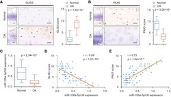
- Figure 5 Expression of GLIS3, PAX5, and miR-106a-5p in OA Patients (A) Representative immunohistochemistry assay of GLIS3 in normal and OA cartilage tissues. Scale bar, left, 500 mum; right, 50 mum. The scores of GLIS3 in normal and OA cartilage tissues based on the immunohistochemistry assay are shown. (B) Representative immunohistochemistry assay of PAX5 in normal and OA cartilage tissues. Scale bar, left, 500 mum; right, 50 mum. The scores of PAX5 in normal and OA cartilage tissues based on the immunohistochemistry assay. (C) qRT-PCR measurement of miR-106a-5p in normal and OA cartilage tissues. Data are expressed as the mean +- SD. (D and E) The relationship among GLIS3 (D), PAX5 (E), and miR-106a-5p expression was determined through Pearson's chi2 analysis.
