 Explore
 Validate
 Learn
Learn Western blot
Antibody data
- Antibody Data
- Antigen structure
- References [0]
- Comments [0]
- Validations
- Western blot [2]
- Immunohistochemistry [1]
- Flow cytometry [1]
Submit
Validation data
Reference
Comment
Report error
- Product number
- MAB3487 - Provider product page

- Provider
- Novus Biologicals
- Product name
- Rabbit Monoclonal Pax5/BSAP Antibody
- Antibody type
- Monoclonal
- Description
- Protein A or G purified from cell culture supernatant. Detects human Pax5 in direct ELISAs and Western blots.
- Reactivity
- Human, Mouse
- Host
- Rabbit
- Conjugate
- Unconjugated
- Isotype
- IgG
- Vial size
- 100 ug
- Storage
- Use a manual defrost freezer and avoid repeated freeze-thaw cycles. 12 months from date of receipt, -20 to -70 degreesC as supplied. 1 month, 2 to 8 degreesC under sterile conditions after reconstitution. 6 months, -20 to -70 degreesC under sterile conditions after reconstitution.
No comments: Submit comment
Supportive validation
- Submitted by
- Novus Biologicals (provider)
- Main image

- Experimental details
- Detection of Human Pax5/BSAP by Western Blot. Western blot shows lysates of Raji human Burkitt's lymphoma cell line, Ramos human Burkitt's lymphoma cell line, Daudi human Burkitt's lymphoma cell line, and Nalm-6 human Pre-B acute lymphocytic leukemia cell line. PVDF membrane was probed with 0.1 µg/mL of Rabbit Anti-Human/Mouse Pax5/BSAP Monoclonal Antibody (Catalog # MAB3487) followed by HRP-conjugated Anti-Rabbit IgG Secondary Antibody (Catalog # HAF008). A specific band was detected for Pax5/BSAP at approximately 42 kDa (as indicated). This experiment was conducted under reducing conditions and using Immunoblot Buffer Group 1.

- Submitted by
- Novus Biologicals (provider)
- Main image

- Experimental details
- Detection of Human Pax5/BSAP by Simple WesternTM. Simple Western lane view shows lysates of Raji human Burkitt's lymphoma cell line, Ramos human Burkitt's lymphoma cell line, Daudi human Burkitt's lymphoma cell line, and Nalm-6 human Pre-B acute lymphocytic leukemia cell line, loaded at 0.2 mg/mL. A specific band was detected for Pax5/BSAP at approximately 56 kDa (as indicated) using 1 µg/mL of Rabbit Anti-Human/Mouse Pax5/BSAP Monoclonal Antibody (Catalog # MAB3487). This experiment was conducted under reducing conditions and using the 12-230 kDa separation system.

Supportive validation
- Submitted by
- Novus Biologicals (provider)
- Main image
- Experimental details
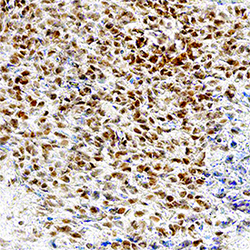
- Pax5/BSAP in Mouse Embryo. Pax5/BSAP was detected in perfusion fixed frozen sections of mouse embryo (13 d.p.c.) using Rabbit Anti-Human/Mouse Pax5/BSAP Monoclonal Antibody (Catalog # MAB3487) at 3 µg/mL overnight at 4 °C. Tissue was stained using the Anti-Rabbit HRP-DAB Cell & Tissue Staining Kit (brown; Catalog # CTS005) and counterstained with hematoxylin (blue). Specific staining was localized to nuclei. View our protocol for Chromogenic IHC Staining of Frozen Tissue Sections.

Supportive validation
- Submitted by
- Novus Biologicals (provider)
- Main image

- Experimental details
- Detection of Pax5/BSAP in Human PBMCs by Flow Cytometry. Human peripheral blood mononuclear cells (PBMCs) were stained with Mouse Anti-Human CD19 APC-conjugated Monoclonal Antibody (Catalog # FAB4867A) and either (A) Rabbit Anti-Human/Mouse Pax5/BSAP Monoclonal Antibody (Catalog # MAB3487) or (B) Normal Rabbit IgG Control (Catalog # MAB1050) followed by Phycoerythrin-conjugated Anti-Rabbit IgG Secondary Antibody (Catalog # F0110). To facilitate intracellular staining, cells were fixed and permeabilized with FlowX FoxP3 Fixation & Permeabilization Buffer Kit (Catalog # FC012). View our protocol for Staining Intracellular Molecules.
