Explore
 Validate
 Learn
Learn Flow cytometry
Antibody data
- Antibody Data
- Antigen structure
- References [2]
- Comments [0]
- Validations
- Flow cytometry [1]
- Other assay [1]
Submit
Validation data
Reference
Comment
Report error
- Product number
- 12-9918-80 - Provider product page

- Provider
- Invitrogen Antibodies
- Product name
- PAX5 Monoclonal Antibody (1H9), PE, eBioscience™
- Antibody type
- Monoclonal
- Antigen
- Other
- Description
- Description: The monoclonal antibody 1H9 recognizes both mouse and human Pax5. Pax5, also known as BSAP (B cell specific activator protein), is a member of the paired box (pax) family of transcription factors. Pax5 is the only member of the pax family of transcription factors that is expressed in hematopoietic cells. During embryogenesis, Pax5 is transiently expressed in the brain of mice and in the mesencephalon and spinal cord of humans. Its expression is upregulated early in B cell development at the time of B cell commitment and is maintained throughout most subsequent stages. Suppression of Pax5 is essential for expression of Blimp-1 and the terminal differentiation of plasma cells. In the spleen, expression of Pax5 is higher in marginal zone B cells (B220+ CD21high CD23low) than in other B cells, especially the transition 1 stage (B220+ CD21- CD23-). In addition to its role in B cell development, Pax5 also affects VH-DJH heavy chain recombination as well as influencing the expression of many other B and non-B cell related proteins. Pax5 expression is correlated with many neoplasms. In diffuse large B cell lymphomas (DLBCL) and non-Hodgkin lymphomas Pax5 is often mutated while in B-cell ALL, expression levels are high. Additionally, translocation with elastin, IGH, ETV6, FOXP1, and EVI3 have been identified.
- Conjugate
- Yellow dye
- Antibody clone number
- 1H9
- Concentration
- 0.2 mg/mL
Submitted references Normal B cell development and Pax5 expression in Thy28/ThyN1-deficient mice.
Reporter gene insertions reveal a strictly B lymphoid-specific expression pattern of Pax5 in support of its B cell identity function.
Kitaura F, Yuno M, Fujita T, Wakana S, Ueda J, Yamagata K, Fujii H
PloS one 2019;14(7):e0220199
PloS one 2019;14(7):e0220199
Reporter gene insertions reveal a strictly B lymphoid-specific expression pattern of Pax5 in support of its B cell identity function.
Fuxa M, Busslinger M
Journal of immunology (Baltimore, Md. : 1950) 2007 Mar 1;178(5):3031-7
Journal of immunology (Baltimore, Md. : 1950) 2007 Mar 1;178(5):3031-7
No comments: Submit comment
Supportive validation
- Submitted by
- Invitrogen Antibodies (provider)
- Main image

- Experimental details
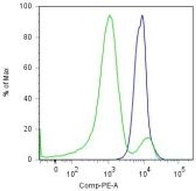
- Surface staining of C57BL/6 splenocytes with Anti-Mouse CD23 PE-Cy7 (Product # 25-0232-82) and Anti-Mouse CD21/CD35 FITC (Product # 11-0211-82) followed by intracellular staining using the Foxp3 Staining Buffer Set (Product # 00-5523-00) and 0.125 µg of Anti-Human/Mouse Pax5 PE. Histogram depicts cell populations based on surface stains comprising the marginal zone cells (CD21 (high)CD23-) (blue histogram) or T1 cells (CD21-CD23-) (green histogram).
- Conjugate
- Yellow dye
Supportive validation
- Submitted by
- Invitrogen Antibodies (provider)
- Main image
- Experimental details
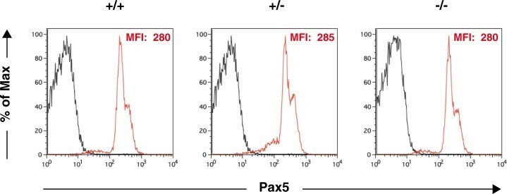
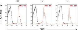
- Fig 7 Expression of Pax5 in splenic B cells. Splenocytes from 7-week-old mice were stained with a FITC-conjugated anti-CD19 Ab and a PE-conjugated anti-Pax5 Ab. The expression level of Pax5 in CD19 + B cells is shown. The mean fluorescence intensity (MFI) of Pax5 staining is shown. Black: unstained control; red: Pax5 staining. Percentages of Pax5 + cells in CD19 + splenic B cells from Thy28 +/ , Thy28 +/- , and Thy28 -/- mice were 95.6%, 94.7%, and 95.6%, respectively.
- Conjugate
- Yellow dye