 Explore
 Validate
 Learn
Learn Flow cytometry
Antibody data
- Antibody Data
- Antigen structure
- References [5]
- Comments [0]
- Validations
- Flow cytometry [1]
- Other assay [2]
Submit
Validation data
Reference
Comment
Report error
- Product number
- 17-9918-80 - Provider product page

- Provider
- Invitrogen Antibodies
- Product name
- PAX5 Monoclonal Antibody (1H9), APC, eBioscience™
- Antibody type
- Monoclonal
- Antigen
- Other
- Description
- Description: The monoclonal antibody 1H9 recognizes both mouse and human Pax5. Pax5, also known as BSAP (B cell specific activator protein), is a member of the paired box (pax) family of transcription factors. Pax5 is the only member of the pax family of transcription factors that is expressed in hematopoietic cells. During embryogenesis, Pax5 is transiently expressed in the brain of mice and in the mesencephalon and spinal cord of humans. Its expression is upregulated early in B cell development at the time of B cell commitment and is maintained throughout most subsequent stages. Suppression of Pax5 is essential for expression of Blimp-1 and the terminal differentiation of plasma cells. In the spleen, expression of Pax5 is higher in marginal zone B cells (B220+ CD21high CD23low) than in other B cells, especially the transition 1 stage (B220+ CD21- CD23-). In addition to its role in B cell development, Pax5 also affects VH-DJH heavy chain recombination as well as influencing the expression of many other B and non-B cell related proteins. Pax5 expression is correlated with many neoplasms. In diffuse large B cell lymphomas (DLBCL) and non-Hodgkin lymphomas Pax5 is often mutated while in B-cell ALL, expression levels are high. Additionally, translocation with elastin, IGH, ETV6, FOXP1, and EVI3 have been identified.
- Antibody clone number
- 1H9
- Concentration
- 0.2 mg/mL
Submitted references The mitochondrial iron transporter ABCB7 is required for B cell development, proliferation, and class switch recombination in mice.
Normal B cell development and Pax5 expression in Thy28/ThyN1-deficient mice.
Differential Effects of Tacrolimus versus Sirolimus on the Proliferation, Activation and Differentiation of Human B Cells.
VprBP Is Required for Efficient Editing and Selection of Igκ+ B Cells, but Is Dispensable for Igλ+ and Marginal Zone B Cell Maturation and Selection.
Reporter gene insertions reveal a strictly B lymphoid-specific expression pattern of Pax5 in support of its B cell identity function.
Lehrke MJ, Shapiro MJ, Rajcula MJ, Kennedy MM, McCue SA, Medina KL, Shapiro VS
eLife 2021 Nov 11;10
eLife 2021 Nov 11;10
Normal B cell development and Pax5 expression in Thy28/ThyN1-deficient mice.
Kitaura F, Yuno M, Fujita T, Wakana S, Ueda J, Yamagata K, Fujii H
PloS one 2019;14(7):e0220199
PloS one 2019;14(7):e0220199
Differential Effects of Tacrolimus versus Sirolimus on the Proliferation, Activation and Differentiation of Human B Cells.
Traitanon O, Mathew JM, La Monica G, Xu L, Mas V, Gallon L
PloS one 2015;10(6):e0129658
PloS one 2015;10(6):e0129658
VprBP Is Required for Efficient Editing and Selection of Igκ+ B Cells, but Is Dispensable for Igλ+ and Marginal Zone B Cell Maturation and Selection.
Palmer VL, Aziz-Seible R, Kassmeier MD, Rothermund M, Perry GA, Swanson PC
Journal of immunology (Baltimore, Md. : 1950) 2015 Aug 15;195(4):1524-37
Journal of immunology (Baltimore, Md. : 1950) 2015 Aug 15;195(4):1524-37
Reporter gene insertions reveal a strictly B lymphoid-specific expression pattern of Pax5 in support of its B cell identity function.
Fuxa M, Busslinger M
Journal of immunology (Baltimore, Md. : 1950) 2007 Mar 1;178(5):3031-7
Journal of immunology (Baltimore, Md. : 1950) 2007 Mar 1;178(5):3031-7
No comments: Submit comment
Supportive validation
- Submitted by
- Invitrogen Antibodies (provider)
- Main image
- Experimental details
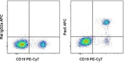
- Intracellular staining of normal human peripheral blood cells with Anti-Human CD19 PE-Cy7 (Product # 25-0199-42) and 0.125 µg of Rat IgG2a K Isotype Control APC (Product # 17-4321-81) (left) or 0.125 µg of Anti-Human/Mouse Pax5 APC (right) using the Foxp3 Staining Buffer Set (Product # 00-5523-00). Cells in the lymphocyte gate were used for analysis.

Supportive validation
- Submitted by
- Invitrogen Antibodies (provider)
- Main image
- Experimental details
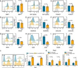
- Figure 2. Gene expression changes confirm absence of pre-B cells in Mb1-cre ABCB7 conditional knockout (cKO) mice. Analysis of critical transcription factors in wild-type (WT) and Mb1-cre ABCB7 cKO Fr. C cells (B220 + CD19 + CD43 + BP-1 + ). ( A-G ) Intracellular flow cytometry analysis of EBF1 ( A ), E47 (E2A) ( B ), FOXO1 ( C ), PAX5 ( D ), IKAROS ( E ), AIOLOS ( F ), and IRF4 ( G ) expression. Quantification of MdFI is shown on the right of each plot. Isotype controls are shown in gray. Offset histograms are representative of at least three independent experiments (total of 6-10 mice/group). ( H, I ) Flow cytometry analysis of CD2 ( H ) and CD25 ( I ) expression. Indicated values are the proportion of Fr. C cells positive for either marker, and quantifications are shown on the right of each plot. Offset histograms are representative of three independent experiments (total of five mice/group). ( J ) Intracellular flow cytometry analysis of TdT expression in Fr. B and Fr. C cells. Indicated values are the proportion of cells positive for TdT expression, and quantifications are shown on the right. Offset histograms are representative of three independent experiments (total of 5-7 mice/group). ( K ) Quantitative real-time PCR analysis of Rag1 and Rag2 expression in sorted Fr. B and Fr. C cells. 18S rRNA was used as an endogenous control, and relative expression values were normalized to expression in WT Fr. B cells. Results were obtained from three independent experiments (tot

- Submitted by
- Invitrogen Antibodies (provider)
- Main image
- Experimental details
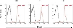
- Fig 7 Expression of Pax5 in splenic B cells. Splenocytes from 7-week-old mice were stained with a FITC-conjugated anti-CD19 Ab and a PE-conjugated anti-Pax5 Ab. The expression level of Pax5 in CD19 + B cells is shown. The mean fluorescence intensity (MFI) of Pax5 staining is shown. Black: unstained control; red: Pax5 staining. Percentages of Pax5 + cells in CD19 + splenic B cells from Thy28 +/ , Thy28 +/- , and Thy28 -/- mice were 95.6%, 94.7%, and 95.6%, respectively.
