Explore
 Validate
 Learn
Learn Western blot
 Immunocytochemistry
Antibody data
- Antibody Data
- Antigen structure
- References [2]
- Comments [0]
- Validations
- Immunocytochemistry [2]
- Immunohistochemistry [3]
- Other assay [2]
Submit
Validation data
Reference
Comment
Report error
- Product number
- PA5-34625 - Provider product page

- Provider
- Invitrogen Antibodies
- Product name
- Phospho-CHK1 (Ser345) Polyclonal Antibody
- Antibody type
- Polyclonal
- Antigen
- Synthetic peptide
- Description
- Recommended positive controls: HCT116, HCT116 cells with 30uM cisplatin treatment for 24hr. Predicted reactivity: Mouse (100%), Rat (100%), Bovine (100%). IHC notes, Requires antigen retrieval using heat mediated 10mM Citrate buffer (pH6.0) or Tris-EDTA buffer (pH8.0) Store product as a concentrated solution. Centrifuge briefly prior to opening the vial.
- Reactivity
- Human, Mouse
- Host
- Rabbit
- Isotype
- IgG
- Vial size
- 100 μL
- Concentration
- 1.66 mg/mL
- Storage
- Store at 4°C short term. For long term storage, store at -20°C, avoiding freeze/thaw cycles.
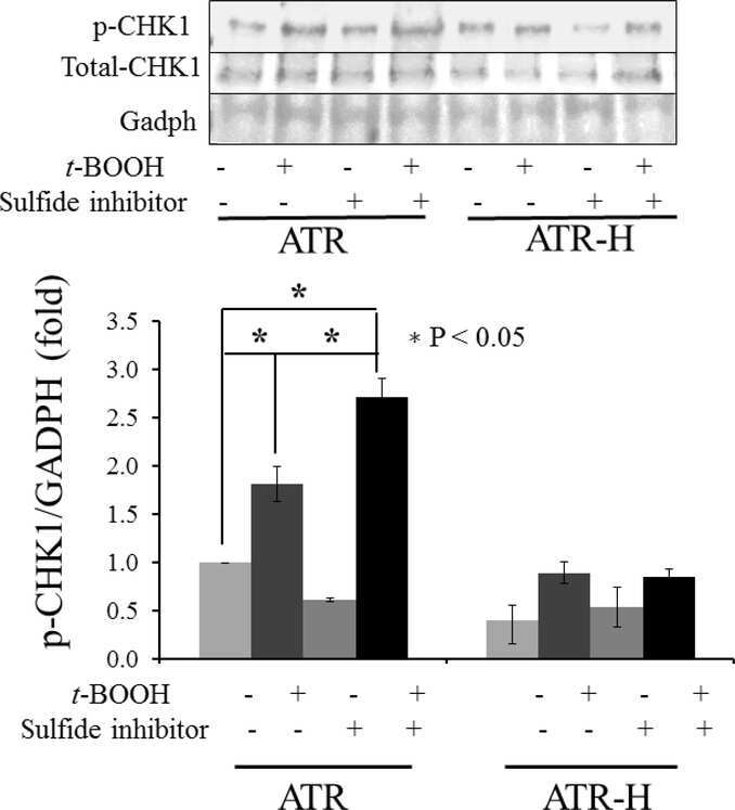
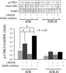
Submitted references The Ataxia telangiectasia-mutated and Rad3-related protein kinase regulates cellular hydrogen sulfide concentrations.
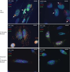
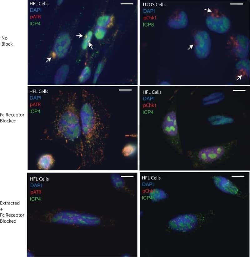
The ATM and Rad3-Related (ATR) Protein Kinase Pathway Is Activated by Herpes Simplex Virus 1 and Required for Efficient Viral Replication.
Chen J, Shen X, Pardue S, Meram AT, Rajendran S, Ghali GE, Kevil CG, Shackelford RE
DNA repair 2019 Jan;73:55-63
DNA repair 2019 Jan;73:55-63
The ATM and Rad3-Related (ATR) Protein Kinase Pathway Is Activated by Herpes Simplex Virus 1 and Required for Efficient Viral Replication.
Edwards TG, Bloom DC, Fisher C
Journal of virology 2018 Mar 15;92(6)
Journal of virology 2018 Mar 15;92(6)
No comments: Submit comment
Supportive validation
- Submitted by
- Invitrogen Antibodies (provider)
- Main image

- Experimental details
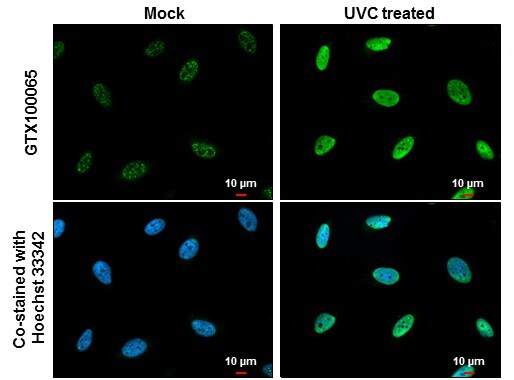
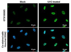
- Immunocytochemistry-Immunofluorescence analysis of Phospho-CHK1 (Ser345) using Phospho-CHK1 (Ser345) Polyclonal Antibody (Product # PA5-34625) (Green) diluted at 1:500. HeLa cells mock (left) and treated with 100 J/m2 UVC and recover for 8 hrs (right) were fixed in 4% paraformaldehyde at RT for 15 min. Blue: Hoechst 33342 staining. Scale bar = 10 µm.
- Submitted by
- Invitrogen Antibodies (provider)
- Main image
- Experimental details
- Immunocytochemistry-Immunofluorescence analysis of Phospho-CHK1 (Ser345) using Phospho-CHK1 (Ser345) Polyclonal Antibody (Product # PA5-34625) (Green) diluted at 1:500. HeLa cells mock (left) and treated with 100 J/m2 UVC and recover for 8 hrs (right) were fixed in 4% paraformaldehyde at RT for 15 min. Blue: Hoechst 33342 staining. Scale bar = 10 µm.

Supportive validation
- Submitted by
- Invitrogen Antibodies (provider)
- Main image
- Experimental details
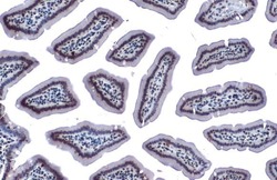
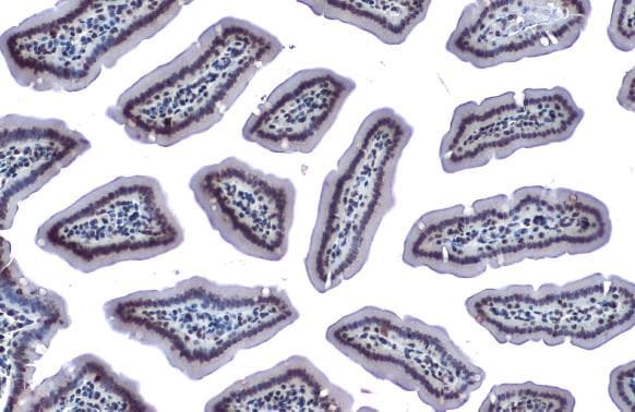
- Phospho-CHK1 (Ser345) Polyclonal Antibody detects Chk1 (phospho Ser345) protein at nucleus by immunohistochemical analysis. Sample: Paraffin-embedded mouse intestine. Chk1 (phospho Ser345) stained by Phospho-CHK1 (Ser345) Polyclonal Antibody (Product # PA5-34625) diluted at 1:500. Antigen Retrieval: Citrate buffer, pH 6.0, 15 min.
- Submitted by
- Invitrogen Antibodies (provider)
- Main image
- Experimental details
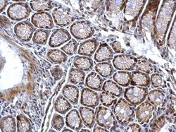
- Phospho-CHK1 (Ser345) Polyclonal Antibody detects Chk1 (phospho Ser345) protein at nucleus on mouse colon by immunohistochemical analysis. Sample: Paraffin-embedded mouse colon. Phospho-CHK1 (Ser345) Polyclonal Antibody (Product # PA5-34625) dilution: 1:500. Antigen Retrieval: EDTA based buffer, pH 8.0, 15 min.

- Submitted by
- Invitrogen Antibodies (provider)
- Main image
- Experimental details
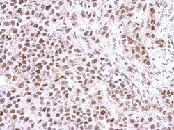
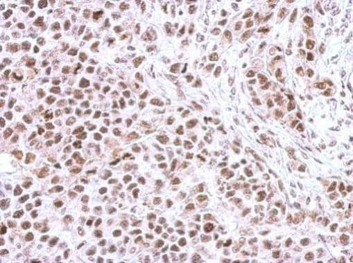
- Immunohistochemical analysis of paraffin-embedded HeLa xenograft, using Chk1 (phospho Ser345) (Product # PA5-34625) antibody at 1:500 dilution. Antigen Retrieval: EDTA based buffer, pH 8.0, 15 min.
Supportive validation
- Submitted by
- Invitrogen Antibodies (provider)
- Main image
- Experimental details
- NULL
- Submitted by
- Invitrogen Antibodies (provider)
- Main image
- Experimental details
- NULL