 Explore
 Validate
 Learn
Learn46-0629-42
antibody from Invitrogen Antibodies
Targeting: SELL
CD62L, hLHRc, LAM-1, LAM1, Leu-8, LNHR, LSEL, Lyam-1, LYAM1, PLNHR
 Flow cytometry
Antibody data
- Antibody Data
- Antigen structure
- References [7]
- Comments [0]
- Validations
- Flow cytometry [1]
- Other assay [7]
Submit
Validation data
Reference
Comment
Report error
- Product number
- 46-0629-42 - Provider product page

- Provider
- Invitrogen Antibodies
- Product name
- CD62L (L-Selectin) Monoclonal Antibody (DREG-56 (DREG56)), PerCP-eFluor™ 710, eBioscience™
- Antibody type
- Monoclonal
- Antigen
- Other
- Description
- Description: The DREG-56 monoclonal antibody reacts with human CD62L, a 76 kDa member of the selectin family. CD62L is expressed by neutrophils, monocytes, and subsets of T, B, and NK cells and binds a number of glycosylated, fucosylated, sulfated sialylated glycoproteins including CD34, glycam-1 and MAdCAM-1. These interactions mediate rolling of lymphocytes on activated endothelium at the sites of inflammation and homing of cells to the high endothelial venules (HEV) of peripheral lymphoid tissues. Applications Reported: This DREG-56 (DREG56) antibody has been reported for use in flow cytometric analysis. Applications Tested: This DREG-56 (DREG56) antibody has been pre-titrated and tested by flow cytometric analysis of normal human peripheral blood cells. This can be used at 5 µL (0.125 µg) per test. A test is defined as the amount (µg) of antibody that will stain a cell sample in a final volume of 100 µL. Cell number should be determined empirically but can range from 10^5 to 10^8 cells/test. PerCP-eFluor® 710 emits at 710 nm and is excited with the blue laser (488 nm); it can be used in place of PerCP-Cyanine5.5. We recommend using a 710/50 bandpass filter, however, the 695/40 bandpass filter is an acceptable alternative. Please make sure that your instrument is capable of detecting this fluorochrome. Fixation: Samples can be stored in IC Fixation Buffer (Product # 00-822-49) (100 µL cell sample + 100 µL IC Fixation Buffer) or 1-step Fix/Lyse Solution (Product # 00-5333-54) for up to 3 days in the dark at 4°C with minimal impact on brightness and FRET efficiency/compensation. Some generalizations regarding fluorophore performance after fixation can be made, but clone specific performance should be determined empirically. Excitation: 488 nm; Emission: 710 nm; Laser: Blue Laser. Filtration: 0.2 µm post-manufacturing filtered.
- Reactivity
- Human
- Host
- Mouse
- Isotype
- IgG
- Antibody clone number
- DREG-56 (DREG56)
- Vial size
- 100 Tests
- Concentration
- 5 µL/Test
- Storage
- 4° C, store in dark, DO NOT FREEZE!
Submitted references Induction of memory-like CD8+ T cells and CD4+ T cells from human naive T cells in culture.
Heterogeneity of human bone marrow and blood natural killer cells defined by single-cell transcriptome.
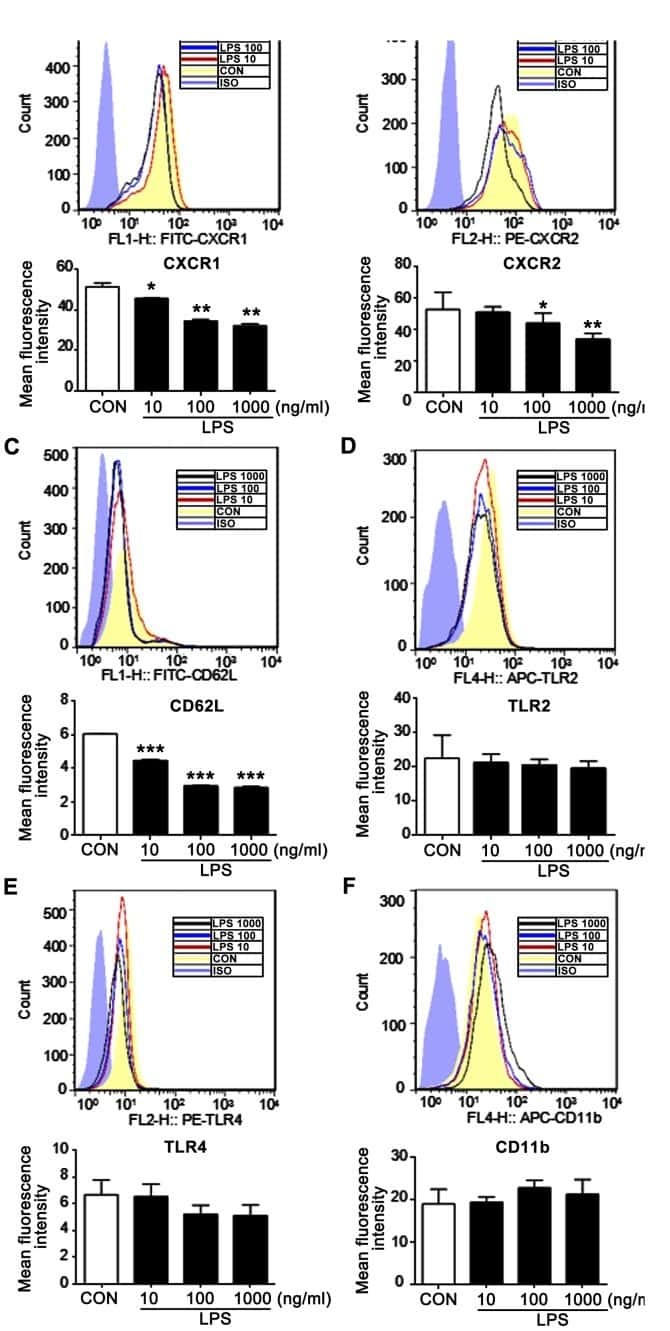
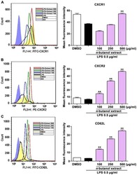
n-butanol extract from Folium isatidis inhibits the lipopolysaccharide-induced downregulation of CXCR1 and CXCR2 on human neutrophils.
Characteristic patterns of HLA presentation and T cell differentiation in adult-onset Still's disease.
Accelerated resolution of inflammation underlies sex differences in inflammatory responses in humans.
L-selectin serves as an E-selectin ligand on cultured human T lymphoblasts.
Identification of a human peripheral lymph node homing receptor: a rapidly down-regulated adhesion molecule.
Tokumoto Y, Araki Y, Narizuka Y, Mizuno Y, Ohshima S, Mimura T
Clinical and experimental immunology 2022 Jan 28;207(1):95-103
Clinical and experimental immunology 2022 Jan 28;207(1):95-103
Heterogeneity of human bone marrow and blood natural killer cells defined by single-cell transcriptome.
Yang C, Siebert JR, Burns R, Gerbec ZJ, Bonacci B, Rymaszewski A, Rau M, Riese MJ, Rao S, Carlson KS, Routes JM, Verbsky JW, Thakar MS, Malarkannan S
Nature communications 2019 Sep 2;10(1):3931
Nature communications 2019 Sep 2;10(1):3931
n-butanol extract from Folium isatidis inhibits the lipopolysaccharide-induced downregulation of CXCR1 and CXCR2 on human neutrophils.
Wu B, Wang L, Jiang L, Dong L, Xu F, Lu Y, Jin J, Wang Z, Liang G, Shan X
Molecular medicine reports 2018 Jan;17(1):179-185
Molecular medicine reports 2018 Jan;17(1):179-185
Characteristic patterns of HLA presentation and T cell differentiation in adult-onset Still's disease.
Jung JY, Choi B, Sayeed HM, Suh CH, Kim YW, Kim HA, Sohn S
International journal of immunopathology and pharmacology 2018 Jan-Dec;32:2058738418791284
International journal of immunopathology and pharmacology 2018 Jan-Dec;32:2058738418791284
Accelerated resolution of inflammation underlies sex differences in inflammatory responses in humans.
Rathod KS, Kapil V, Velmurugan S, Khambata RS, Siddique U, Khan S, Van Eijl S, Gee LC, Bansal J, Pitrola K, Shaw C, D'Acquisto F, Colas RA, Marelli-Berg F, Dalli J, Ahluwalia A
The Journal of clinical investigation 2017 Jan 3;127(1):169-182
The Journal of clinical investigation 2017 Jan 3;127(1):169-182
L-selectin serves as an E-selectin ligand on cultured human T lymphoblasts.
Jutila MA, Kurk S, Jackiw L, Knibbs RN, Stoolman LM
Journal of immunology (Baltimore, Md. : 1950) 2002 Aug 15;169(4):1768-73
Journal of immunology (Baltimore, Md. : 1950) 2002 Aug 15;169(4):1768-73
Identification of a human peripheral lymph node homing receptor: a rapidly down-regulated adhesion molecule.
Kishimoto TK, Jutila MA, Butcher EC
Proceedings of the National Academy of Sciences of the United States of America 1990 Mar;87(6):2244-8
Proceedings of the National Academy of Sciences of the United States of America 1990 Mar;87(6):2244-8
No comments: Submit comment
Supportive validation
- Submitted by
- Invitrogen Antibodies (provider)
- Main image

- Experimental details
- Staining of normal human peripheral blood cells with Anti-Human CD4 APC (Product # 17-0048-42) and Mouse IgG1 K Isotype Control PerCP-eFluor® 710 (Product # 46-4714-82) (left) or Anti-Human CD62L (L-Selectin) PerCP-eFluor® 710 (right). Cells in the lymphocyte gate were used for analysis.

Supportive validation
- Submitted by
- Invitrogen Antibodies (provider)
- Main image
- Experimental details
- NULL
- Submitted by
- Invitrogen Antibodies (provider)
- Main image
- Experimental details
- NULL
- Submitted by
- Invitrogen Antibodies (provider)
- Main image
- Experimental details
- NULL
- Submitted by
- Invitrogen Antibodies (provider)
- Main image
- Experimental details
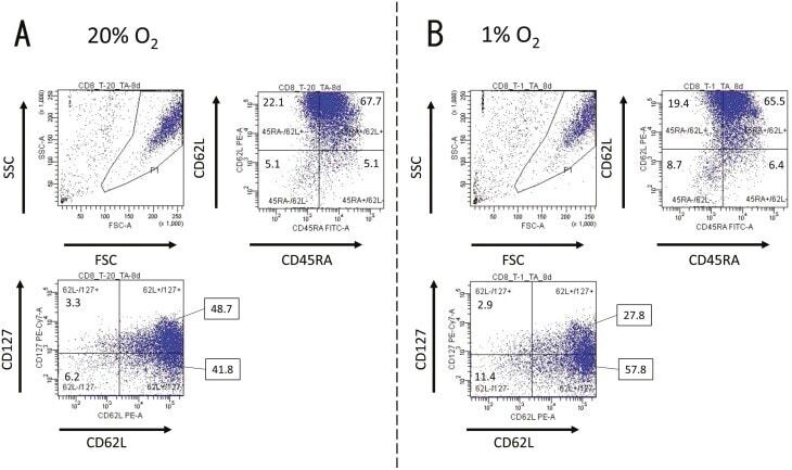
- Fig. 1. Activated CD8 + T cells in either normal or hypoxic culture. Naive CD8 + T cells derived from a healthy donor were cultured in human T-activator CD3/CD28 and IL-2 containing medium for 8 days. ( A ) in 20% O 2 condition and ( B ) in 1% O 2 condition. The 7-AAD-negative cells in the area gated as P1 in the FSC/SSC panel were considered as living cells. The expression pattern of CD45RA, CD62L, and CD127 of living cells were analyzed by FACS. The numbers on the FACS-plot panel mean the frequencies (%) of population of cells. We repeated this experiment four times.
- Submitted by
- Invitrogen Antibodies (provider)
- Main image
- Experimental details
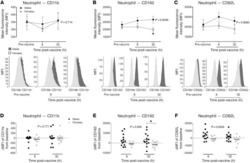
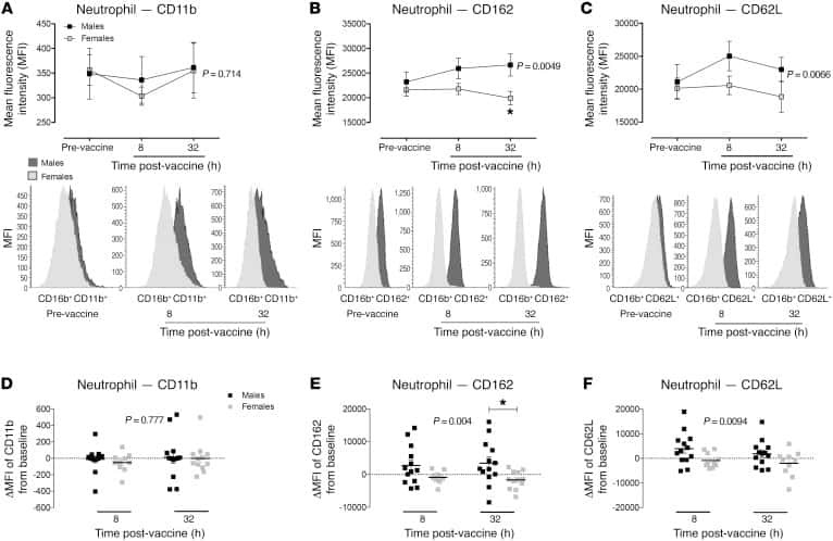
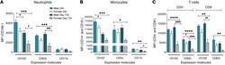
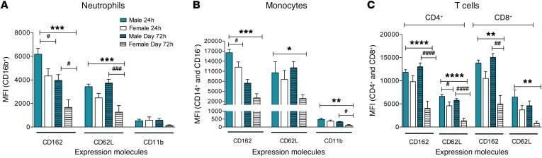
- Figure 5 Reduced inflammatory cell activation state in cantharidin-induced blister exudates in female compared with male healthy volunteers. Mean fluorescence intensity (MFI) of the expression molecules CD162, CD62L, and CD11b on ( A ) neutrophils, ( B ) inflammatory monocytes, and ( C ) CD4 + and CD8 + T cells in healthy male ( n = 16) and female ( n = 16) volunteers. Data are shown as mean +- SEM. Statistical significances determined using 2-way ANOVA, * P < 0.05, ** P < 0.01, *** P < 0.001, and **** P < 0.0001; followed by Sidak's post tests, # P < 0.05, ## P < 0.01, and #### P < 0.0001 comparing the sexes at each time point for all the panels.
- Submitted by
- Invitrogen Antibodies (provider)
- Main image
- Experimental details
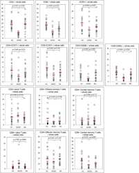
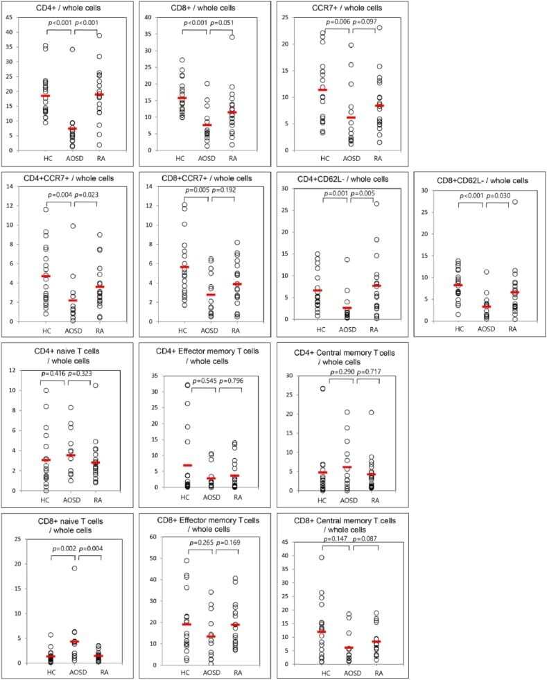
- Figure 2. Percentage of surface-stained cells presenting CD4+, CD8+, CCR7+, CD4+CCR7+, CD8+CCR7+, CD4+CD62L-, CD8+CD62L-, CD4+naive T cell, CD4+ effector memory T cell, CD4+ central memory T cells, CD8+ naive T cells, CD8+ effector memory T cells, and CD8+ central memory T cells in patients with AOSD, a patient with rheumatoid arthritis (RA), and a healthy control (HC). Results were obtained from 14 patients with AOSD, 20 RA patients, and 20 HCs. The horizontal line indicates the mean value for each group. The P value was determined by the Mann-Whitney U test.
- Submitted by
- Invitrogen Antibodies (provider)
- Main image

- Experimental details
- Fig. 4 Active NK cells with a unique transcriptome profile. a Top two enriched gene sets (ranked by normalized enrichment score) of five different datasets from GSEA of the ""Inflamed NK"" cluster compared to the rest of the cells were plotted. b The expression of CD69 in the BM sample was shown as a violin plot. The y -axis represents log-normalized expression value. c Module score was calculated using up-regulated DEGs of ""Active NK"" (left) or ""Inflamed NK"" (right) cluster from BM sample and plotted via violin plots. d Up-regulated IEGs from ""Active NK"" cluster were plotted using heatmap of the BM sample. e The expression of CXCR4 in the BM sample was shown as a violin plot. The y -axis represents log-normalized expression value. f Percentage of CXCR4 + NK cells (gated on Lin - CD56 + cells) was evaluated via flow cytometry. g The expression of CXCR4 in CD57 +/- , CD62L +/- , or NKG2A +/- CD56 dim NK populations from BM was assessed via flow cytometry (top). Percentage of CXCR4 + cells within each population were quantified (bottom). n >= 6 from two to five independent experiments. Paired Student's t test was used for the statistical analysis. * P < 0.05; ** p < 0.01; *** p < 0.001; n.s. stands for ""not significant."" Source data for f and g are provided as a Source Data file. See also Supplementary Fig. 5
