 Explore
 Validate
 Learn
Learn Western blot
 Immunoprecipitation
Antibody data
- Antibody Data
- Antigen structure
- References [0]
- Comments [0]
- Validations
- Western blot [1]
- Immunohistochemistry [4]
Submit
Validation data
Reference
Comment
Report error
- Product number
- LS-C148268 - Provider product page

- Provider
- LSBio
- Product name
- FAP-1 / PTPN13 Antibody (aa1279-1883) LS-C148268
- Antibody type
- Polyclonal
- Description
- Antiserum
- Reactivity
- Human
- Host
- Rabbit
- Storage
- Store at -20°C. Aliquot to avoid freeze/thaw cycles.
No comments: Submit comment
Enhanced validation
- Submitted by
- LSBio (provider)
- Enhanced method
- Genetic validation
- Main image
- Experimental details
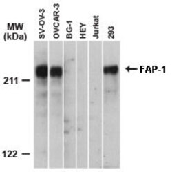
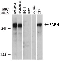
- Western blot of FAP-1 using Polyclonal Antibody to FAP-1 at 1:2000. In ovarian carcinoma cell lines FAP-1 expression was detected in SK-OV-3 and OVCAR-3, but not in BG-1 or HEY. Human Jurkat T and 293 kidney cell lines were used as negative and positive controls, respectively.
Supportive validation
- Submitted by
- LSBio (provider)
- Enhanced method
- Genetic validation
- Main image

- Experimental details
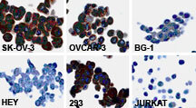
- Immunocytochemistry of FAP-1 in cell lines using Polyclonal Antibody to FAP-1 at 1:2000. In ovarian carcinoma cell lines FAP-1 expression was detected in SK-OV-3 and OVCAR-3, but not in BG-1 or HEY. Human 293 kidney and Jurkat T cell lines were used as positive and negative controls, respectively. The staining data correlates with the western blot data (figure to the left).

- Submitted by
- LSBio (provider)
- Enhanced method
- Genetic validation
- Main image
- Experimental details
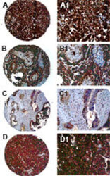
- Immunohistochemistry of FAP-1 in formalin-fixed, paraffin embedded ovarian carcinoma cores from a tissue microarray using Polyclonal Antibody to FAP-1 at 1:2000. A-D, samples are from four different patients. A1-D1 are high magnification images from A-D, respectively. Hematoxylin-Eosin counterstain.

- Submitted by
- LSBio (provider)
- Main image

- Experimental details
- Immunocytochemistry of FAP-1 in cell lines using Polyclonal Antibody to FAP-1 at 1:2000. In ovarian carcinoma cell lines FAP-1 expression was detected in SK-OV-3 and OVCAR-3, but not in BG-1 or HEY. Human 293 kidney and Jurkat T cell lines were used as positive and negative controls, respectively. The staining data correlates with the western blot data (figure to the left).
- Submitted by
- LSBio (provider)
- Main image

- Experimental details
- Immunohistochemistry of FAP-1 in formalin-fixed, paraffin embedded ovarian carcinoma cores from a tissue microarray using Polyclonal Antibody to FAP-1 at 1:2000. A-D, samples are from four different patients. A1-D1 are high magnification images from A-D, respectively. Hematoxylin-Eosin counterstain.
