 Explore
 Validate
 Learn
Learn Western blot
Antibody data
- Antibody Data
- Antigen structure
- References [3]
- Comments [0]
- Validations
- Western blot [8]
- Immunocytochemistry [2]
- Immunohistochemistry [7]
Submit
Validation data
Reference
Comment
Report error
- Product number
- GTX112980 - Provider product page

- Provider
- GeneTex
- Proper citation
- GeneTex Cat#GTX112980, RRID:AB_1951939
- Product name
- SMAD4 antibody
- Antibody type
- Polyclonal
- Reactivity
- Human, Mouse, Rat
- Host
- Rabbit
Submitted references Long-term growth comparison studies of FBS and FBS alternatives in six head and neck cell lines.
TGF-β1 secreted by Tregs in lymph nodes promotes breast cancer malignancy via up-regulation of IL-17RB.
Candesartan ameliorates arsenic-induced hypertensive vascular remodeling by regularizing angiotensin II and TGF-beta signaling in rats.
Fang CY, Wu CC, Fang CL, Chen WY, Chen CL
PloS one 2017;12(6):e0178960
PloS one 2017;12(6):e0178960
TGF-β1 secreted by Tregs in lymph nodes promotes breast cancer malignancy via up-regulation of IL-17RB.
Huang SC, Wei PC, Hwang-Verslues WW, Kuo WH, Jeng YM, Hu CM, Shew JY, Huang CS, Chang KJ, Lee EY, Lee WH
EMBO molecular medicine 2017 Dec;9(12):1660-1680
EMBO molecular medicine 2017 Dec;9(12):1660-1680
Candesartan ameliorates arsenic-induced hypertensive vascular remodeling by regularizing angiotensin II and TGF-beta signaling in rats.
Khuman MW, Harikumar SK, Sadam A, Kesavan M, Susanth VS, Parida S, Singh KP, Sarkar SN
Toxicology 2016 Dec 30;374:29-41
Toxicology 2016 Dec 30;374:29-41
No comments: Submit comment
Supportive validation
- Submitted by
- GeneTex (provider)
- Main image

- Experimental details
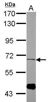
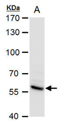
- Sample (50 ug of whole cell lysate) A: mouse muscle 7.5% SDS PAGE GTX112980 diluted at 1:1000
- Validation comment
- WB
- Submitted by
- GeneTex (provider)
- Main image
- Experimental details
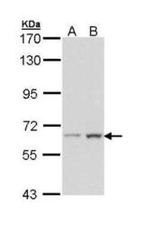
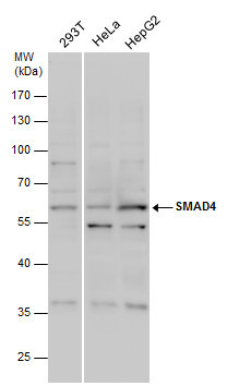
- Sample (30 ug of whole cell lysate) A: Hela B: Hep G2 (GTX27900) 7.5% SDS PAGE GTX112980 diluted at 1:1000
- Validation comment
- WB
- Submitted by
- GeneTex (provider)
- Main image
- Experimental details
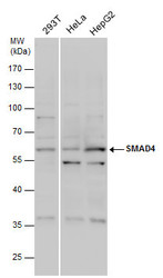
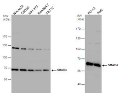
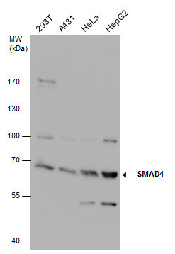
- SMAD4 antibody detects SMAD4 protein by western blot analysis. Various whole cell extracts (30 ?g) were separated by 10% SDS-PAGE, and the membrane was blotted with SMAD4 antibody (GTX112980) diluted by 1:1000.
- Validation comment
- WB
- Submitted by
- GeneTex (provider)
- Main image
- Experimental details
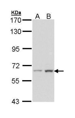
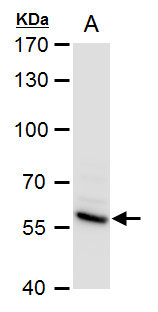
- SMAD4 antibody detects SMAD4 protein by western blot analysis.A. 30 ?g PC-12 whole cell extract 7.5 % SDS-PAGESMAD4 antibody (GTX112980) dilution: 1:1000
- Validation comment
- WB
- Submitted by
- GeneTex (provider)
- Main image
- Experimental details
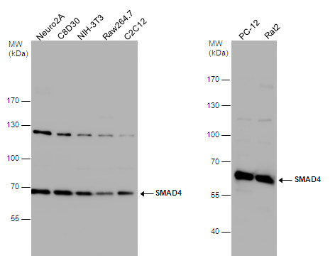
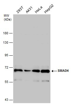
- SMAD4 antibody detects SMAD4 protein by western blot analysis. Various whole cell extracts were separated by 7.5% SDS-PAGE, and the membrane was blotted with SMAD4 antibody (GTX112980) diluted at 1:1000. The HRP-conjugated anti-rabbit IgG antibody (GTX213110-01) was used to detect the primary antibody.
- Submitted by
- GeneTex (provider)
- Main image
- Experimental details
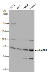
- SMAD4 antibody detects SMAD4 protein by western blot analysis. Various whole cell extracts (30 ?g) were separated by 7.5% SDS-PAGE, and the membrane was blotted with SMAD4 antibody (GTX112980) diluted at 1:1000.
- Validation comment
- WB
- Submitted by
- GeneTex (provider)
- Main image
- Experimental details
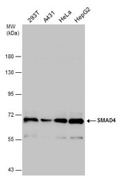
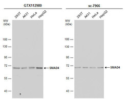
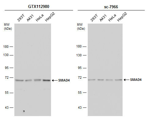
- Various whole cell extracts (30 ?g) were separated by 7.5% SDS-PAGE, and the membrane was blotted with SMAD4 antibody (GTX112980) diluted at 1:1000. The HRP-conjugated anti-rabbit IgG antibody (GTX213110-01) was used to detect the primary antibody.
- Submitted by
- GeneTex (provider)
- Main image
- Experimental details
- Various whole cell extracts (30 ?g) were separated by 7.5% SDS-PAGE, and the membranes were blotted with SMAD4 antibody (GTX112980) diluted at 1:1000 and competitor's antibody (sc-7966) diluted at 1:100. The HRP-conjugated anti-rabbit IgG antibody (GTX213110-01) was used to detect the primary antibody.
Supportive validation
- Submitted by
- GeneTex (provider)
- Main image

- Experimental details
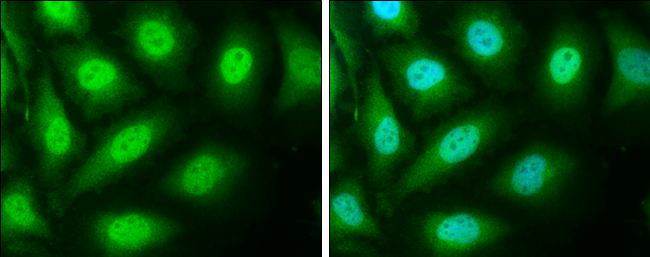
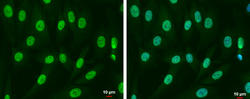
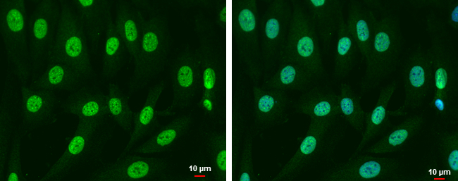
- SMAD4 antibody detects SMAD4 protein at cytoplasm and nucleus by immunofluorescent analysis.Sample: HeLa cells were fixed in 4% paraformaldehyde at RT for 15 min.Green: SMAD4 protein stained by SMAD4 antibody (GTX112980) diluted at 1:500.Blue: Hoechst 33342 staining.
- Submitted by
- GeneTex (provider)
- Main image
- Experimental details
- SMAD4 antibody detects SMAD4 protein a tcytoplasm and nucleus by immunofluorescent analysis.Sample: SK-N-SH cells were fixed in 4% paraformaldehyde at RT for 15 min.Green: SMAD4 protein stained by SMAD4 antibody (GTX112980) diluted at 1:500.Blue: Hoechst 33342 staining.Scale bar = 10 £gm.
Supportive validation
- Submitted by
- GeneTex (provider)
- Main image
- Experimental details
- Immunohistochemical analysis of paraffin-embedded human ulcerative colitis tissue using SMAD4 antibody (GTX112980)
- Submitted by
- GeneTex (provider)
- Main image
- Experimental details
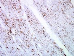
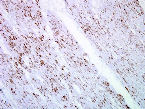
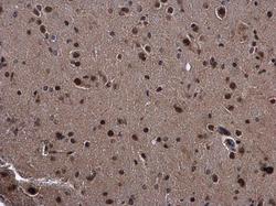
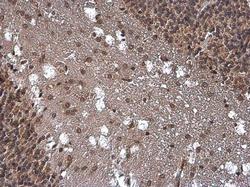
- SMAD4 antibody detects SMAD4 protein at cytoplasm and nucleus in mouse brain by immunohistochemical analysis. Sample: Paraffin-embedded mouse brain. SMAD4 antibody (GTX112980) diluted at 1:500.
- Submitted by
- GeneTex (provider)
- Main image
- Experimental details
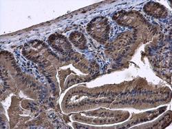
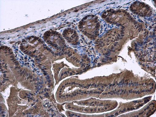
- SMAD4 antibody detects SMAD4 protein at cytoplasm and nucleus in mouse duodenum by immunohistochemical analysis. Sample: Paraffin-embedded mouse duodenum. SMAD4 antibody (GTX112980) diluted at 1:500.
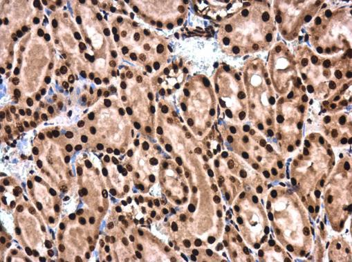
- Submitted by
- GeneTex (provider)
- Main image
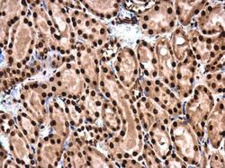
- Experimental details
- SMAD4 antibody detects SMAD4 protein at cytoplasm and nucleus in rat kidney by immunohistochemical analysis. Sample: Paraffin-embedded rat kidney. SMAD4 antibody (GTX112980) diluted at 1:500.
- Submitted by
- GeneTex (provider)
- Main image
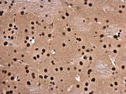
- Experimental details
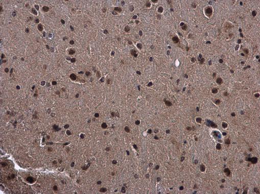
- SMAD4 antibody detects SMAD4 protein at cytoplasm and nucleus in rat brain by immunohistochemical analysis. Sample: Paraffin-embedded rat brain. SMAD4 antibody (GTX112980) diluted at 1:500.
- Submitted by
- GeneTex (provider)
- Main image
- Experimental details
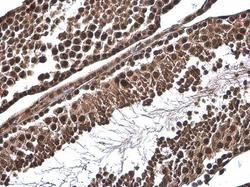
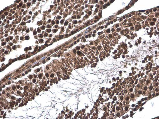
- SMAD4 antibody detects SMAD4 protein at cytoplasm and nucleus by immunohistochemical analysis.Sample: Paraffin-embedded mouse testis.SMAD4 stained by SMAD4 antibody (GTX112980) diluted at 1:500.
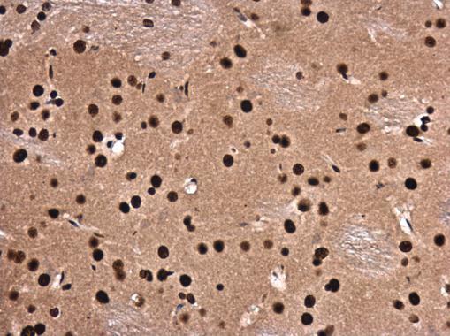
- Submitted by
- GeneTex (provider)
- Main image
- Experimental details
- SMAD4 antibody detects SMAD4 protein at cytoplasm and nucleus by immunohistochemical analysis.Sample: Paraffin-embedded rat brain.SMAD4 stained by SMAD4 antibody (GTX112980) diluted at 1:500.
