Explore
 Validate
 Learn
Learn Flow cytometry
Antibody data
- Antibody Data
- Antigen structure
- References [6]
- Comments [0]
- Validations
- Flow cytometry [2]
- Other assay [1]
Submit
Validation data
Reference
Comment
Report error
- Product number
- Q10065 - Provider product page

- Provider
- Invitrogen Antibodies
- Product name
- CD27 Monoclonal Antibody (CLB-27/1), Qdot™ 605
- Antibody type
- Monoclonal
- Antigen
- Other
- Description
- Qdot Antibody (Ab) conjugates possess a bright fluorescence emission that makes them well suited for the detection of low-abundance extracellular proteins. Approximately the same size as R-phycoerythrin (R-PE) and compatible with existing organic fluorophore conjugates, Qdot Ab conjugates can be excited with any wavelength below their emission maximum, but are best excited by UV or violet light. The narrow, symmetric emission profiles of Qdot Ab conjugates allow for minimal compensation when using a single excitation source, and the very long stoke shifts enable better, more efficient multicolor assays using the 405 nm violet laser. Available in multiple colors for use in flow cytometry, these advantages make Qdot Ab conjugates powerful tools for antibody labeling and staining. Staining: Stain cells in any standard staining buffer, such as phosphate buffered saline (PBS) with 1% bovine serum albumin (BSA). Use 1 L of Qdot antibody conjugate per 1 × 10^6 cells in a 100 µL staining volume (20 nM final concentration of Qdot nanocrystals). Qdot nanocrystal conjugates may be mixed with other antibodies, but use the diluted conjugates on the day of dilution. Qdot nanocrystal conjugates can be used for surface staining applications with most conventional sample preparation reagents, such as Cal-Lyse and FIX & PERM reagents, with minimal affect on fluorescence. We have observed that some batches of BD FACS Lysing Solution have been found to interfere with Qdot nanocrystal fluorescence. Each lot has been tested by flow cytometry using human peripheral blood leukocytes. This testing was performed using 1 L of antibody per 1 × 10⁶ cells in a 100 L staining volume (20 nM final concentration of Qdot nanocrystals). Qdot nanocrystal concentration is assigned based on optical density. The isotype control for this antibody is mouse IgG2a, Cat. No. Q10014. Instrument setup: Qdot Ab conjugates are excited optimally with UV or 405 nm light, although excitation can be obtained with any wavelength below the emission maximum of a given Qdot nanocrystal, such as with a 488-nm laser. Qdot Ab conjugates can be used on cytometers that do not have UV or violet excitation sources as long as they have appropriate emission filters. Make sure the cytometer has an appropriate emission filter for the Qdot Ab conjugate being used; alternate filters can be used as long as they capture the emission maximum, but filter width impacts spectral overlap corrections. And be sure to check for Qdot Ab conjugate emission in any channel that can capture nanocrystal emission off of other lasers on the cytometer. For Cat. No. Q10065: peak excitation 405 (488) nm/peak emission 605 nm; recommended filter 605/20 nm. Store reagents at 2-8°C in the dark. Do not freeze. Because Qdot nanocrystals are conjugated to biological materials, some loss of activity may be observed with prolonged storage. When stored as instructed, expires six months from date of receipt unless otherwise indicated on product label. Qdot Ab conjugates are photostable, and do not need to be protected from light. However, if using Qdot Ab conjugates in combination with conventional fluorochrome conjugated antibodies, minimize light exposure during handling, incubation with cells, and prior to analysis. The Qdot Ab conjugates contain cadmium and selenium in an inorganic crystalline form. Dispose of the material in compliance with all applicable local, state, and federal regulations for disposal of these classes of material. For more information on the composition of these materials, consult the Safety Data Sheets (SDSs).
- Reactivity
- Human
- Host
- Mouse
- Isotype
- IgG
- Antibody clone number
- CLB-27/1
- Vial size
- 100 μL
- Storage
- 4°C, store in dark
Submitted references CD57(+) Memory T Cells Proliferate In Vivo.
Selective Loss of Early Differentiated, Highly Functional PD1high CD4 T Cells with HIV Progression.
Longitudinal studies of a B cell-derived signature of tolerance in renal transplant recipients.
Analysis of human B-cell responses following ChAd63-MVA MSP1 and AMA1 immunization and controlled malaria infection.
Multiparameter flow cytometry and bioanalytics for B cell profiling in systemic lupus erythematosus.
Decreased influenza-specific B cell responses in rheumatoid arthritis patients treated with anti-tumor necrosis factor.
Ahmed R, Miners KL, Lahoz-Beneytez J, Jones RE, Roger L, Baboonian C, Zhang Y, Wang ECY, Hellerstein MK, McCune JM, Baird DM, Price DA, Macallan DC, Asquith B, Ladell K
Cell reports 2020 Dec 15;33(11):108501
Cell reports 2020 Dec 15;33(11):108501
Selective Loss of Early Differentiated, Highly Functional PD1high CD4 T Cells with HIV Progression.
Paris RM, Petrovas C, Ferrando-Martinez S, Moysi E, Boswell KL, Archer E, Yamamoto T, Ambrozak D, Casazza JP, Haubrich R, Connors M, Ake J, Kim JH, Koup RA
PloS one 2015;10(12):e0144767
PloS one 2015;10(12):e0144767
Longitudinal studies of a B cell-derived signature of tolerance in renal transplant recipients.
Newell KA, Asare A, Sanz I, Wei C, Rosenberg A, Gao Z, Kanaparthi S, Asare S, Lim N, Stahly M, Howell M, Knechtle S, Kirk A, Marks WH, Kawai T, Spitzer T, Tolkoff-Rubin N, Sykes M, Sachs DH, Cosimi AB, Burlingham WJ, Phippard D, Turka LA
American journal of transplantation : official journal of the American Society of Transplantation and the American Society of Transplant Surgeons 2015 Nov;15(11):2908-20
American journal of transplantation : official journal of the American Society of Transplantation and the American Society of Transplant Surgeons 2015 Nov;15(11):2908-20
Analysis of human B-cell responses following ChAd63-MVA MSP1 and AMA1 immunization and controlled malaria infection.
Elias SC, Choudhary P, de Cassan SC, Biswas S, Collins KA, Halstead FD, Bliss CM, Ewer KJ, Hodgson SH, Duncan CJ, Hill AV, Draper SJ
Immunology 2014 Apr;141(4):628-44
Immunology 2014 Apr;141(4):628-44
Multiparameter flow cytometry and bioanalytics for B cell profiling in systemic lupus erythematosus.
Kaminski DA, Wei C, Rosenberg AF, Lee FE, Sanz I
Methods in molecular biology (Clifton, N.J.) 2012;900:109-34
Methods in molecular biology (Clifton, N.J.) 2012;900:109-34
Decreased influenza-specific B cell responses in rheumatoid arthritis patients treated with anti-tumor necrosis factor.
Kobie JJ, Zheng B, Bryk P, Barnes M, Ritchlin CT, Tabechian DA, Anandarajah AP, Looney RJ, Thiele RG, Anolik JH, Coca A, Wei C, Rosenberg AF, Feng C, Treanor JJ, Lee FE, Sanz I
Arthritis research & therapy 2011;13(6):R209
Arthritis research & therapy 2011;13(6):R209
No comments: Submit comment
Supportive validation
- Submitted by
- Invitrogen Antibodies (provider)
- Main image
- Experimental details
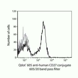
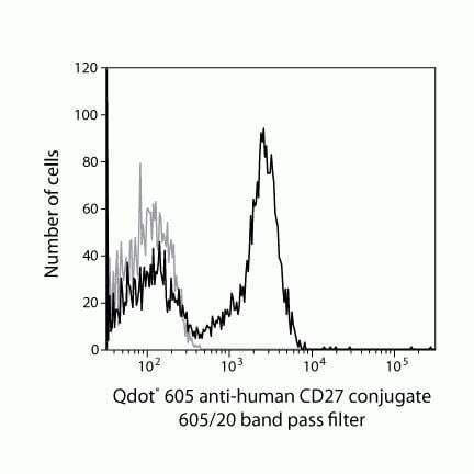
- Qdot® 605 anti-human CD27 conjugate 605/20 band pass filter

- Submitted by
- Invitrogen Antibodies (provider)
- Main image

- Experimental details
- Qdot® 605 anti-human CD27 conjugate 605/20 band pass filter
Supportive validation
- Submitted by
- Invitrogen Antibodies (provider)
- Main image
- Experimental details
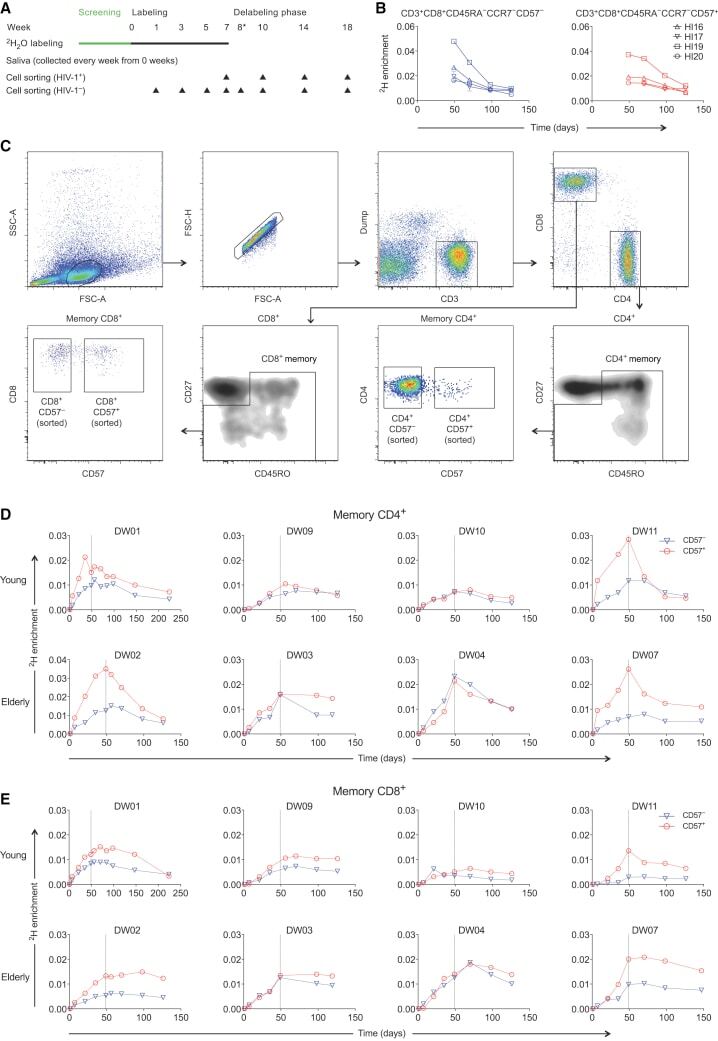
- Figure 1 CD57 - and CD57 + Memory T Cells Exhibit Similar Rates of Deuterium Incorporation (A) Schematic representation of the 2 H 2 O labeling protocol and sampling time points. (B) Experimental labeling data for CD57 - and CD57 + memory CD8 + T cells sampled from the HIV-1-infected volunteers in cohort 1. The corresponding flow cytometric gating strategy is shown in Figure S1 . (C) Successive panels depict the flow cytometric gating strategy used to sort CD57 - and CD57 + memory T cells from the CD4 + and CD8 + lineages (cohort 2). Lymphocytes were identified in a forward scatter-area versus side scatter-area plot, and single cells were identified in a forward scatter-area versus forward scatter-height plot. Boolean gates were drawn for analysis only to exclude fluorochrome aggregates. Viable CD3 + CD14 - CD19 - cells were then identified in the CD4 + and CD8 + lineages, and sort gates were fixed on CD57 - and CD57 + memory cells after exclusion of potentially naive CD27 bright CD45RO - cells. (D) Experimental labeling data for CD57 - and CD57 + memory CD4 + T cells sampled from the healthy volunteers in cohort 2. (E) Experimental labeling data for CD57 - and CD57 + memory CD8 + T cells sampled from the healthy volunteers in cohort 2.