 Explore
 Validate
 Learn
Learn Western blot
Antibody data
- Antibody Data
- Antigen structure
- References [3]
- Comments [0]
- Validations
- Western blot [2]
- Immunocytochemistry [1]
- Flow cytometry [1]
- Other assay [3]
Submit
Validation data
Reference
Comment
Report error
- Product number
- MA1-198 - Provider product page

- Provider
- Invitrogen Antibodies
- Product name
- VE-cadherin Monoclonal Antibody (BV9)
- Antibody type
- Monoclonal
- Antigen
- Recombinant full-length protein
- Description
- MA1-198 VE-Cadherin antibody detects human VE-cadherin by western blot, immunofluorescence and flow cytometry. This monoclonal antibody recognizes the extracellular domain of the VE-cadherin protein which is highly expressed at intercellular junctions of vascular endothelial cells such as Huvec.
- Reactivity
- Human
- Host
- Mouse
- Isotype
- IgG
- Antibody clone number
- BV9
- Vial size
- 100 µg
- Concentration
- 1 mg/mL
- Storage
- -20° C, Avoid Freeze/Thaw Cycles
Submitted references Changes in Human Foetal Osteoblasts Exposed to the Random Positioning Machine and Bone Construct Tissue Engineering.
Microgravity Affects Thyroid Cancer Cells during the TEXUS-53 Mission Stronger than Hypergravity.
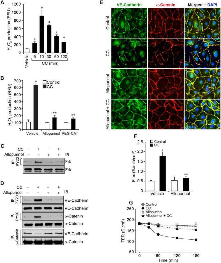
Cholesterol crystals increase vascular permeability by inactivating SHP2 and disrupting adherens junctions.
Mann V, Grimm D, Corydon TJ, Krüger M, Wehland M, Riwaldt S, Sahana J, Kopp S, Bauer J, Reseland JE, Infanger M, Mari Lian A, Okoro E, Sundaresan A
International journal of molecular sciences 2019 Mar 18;20(6)
International journal of molecular sciences 2019 Mar 18;20(6)
Microgravity Affects Thyroid Cancer Cells during the TEXUS-53 Mission Stronger than Hypergravity.
Kopp S, Krüger M, Bauer J, Wehland M, Corydon TJ, Sahana J, Nassef MZ, Melnik D, Bauer TJ, Schulz H, Schütte A, Schmitz B, Oltmann H, Feldmann S, Infanger M, Grimm D
International journal of molecular sciences 2018 Dec 12;19(12)
International journal of molecular sciences 2018 Dec 12;19(12)
Cholesterol crystals increase vascular permeability by inactivating SHP2 and disrupting adherens junctions.
Mani AM, Chattopadhyay R, Singh NK, Rao GN
Free radical biology & medicine 2018 Aug 1;123:72-84
Free radical biology & medicine 2018 Aug 1;123:72-84
No comments: Submit comment
Supportive validation
- Submitted by
- Invitrogen Antibodies (provider)
- Main image
- Experimental details
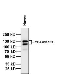
- Western blot analysis of VE-Cadherin was performed by loading 20 µg of huvec whole cell lysate and 5 µL of PageRuler Plus Prestained Protein Ladder (Product # 26619) per well onto a 4-20% Tris-Glycine polyacrylamide gel (Product # WT4202BX10). Proteins were transferred to a nitrocellulose membrane using the G2 Blotter (Product # 62288), and blocked with 5% Milk in TBST for 1 hour at room temperature. VE-Cadherin was detected at ~130-140 kDa using a VE-Cadherin mouse monoclonal antibody (Product # MA1-198) at a dilution of 1:500 in blocking buffer overnight at 4°C on a rocking platform, followed by a Goat anti-Mouse IgG (H+L) Superclonal™ Secondary Antibody, HRP conjugate (Product # A28177) at a dilution of 1:1000 for at least 30 minutes at room temperature. Chemiluminescent detection was performed using SuperSignal West Pico (Product # 34078).

- Submitted by
- Invitrogen Antibodies (provider)
- Main image

- Experimental details
- Western blot was performed using Anti-VE-cadherin Monoclonal Antibody (BV9) (Product # MA1-198) and ~80,100,130 kDa bands corresponding to Cadherin-5 and its glycosylated forms, were observed across high expressing cell lines (HUVEC and BeWo) tested, and not in low expressing cell lines (MCF7, OVCAR3, HeLa and Hep G2). Whole extracts (Scraped) (30 µg lysate) of HUVEC (Lane 1), MCF7 (Lane 2), OVCAR3 (Lane 3), HeLa (Lane 4), Hep G2 (Lane 5) and BeWo (Lane 6) were electrophoresed using NuPAGE™ 10% Bis-Tris Protein Gel (Product # NP0302BOX). Resolved proteins were then transferred onto a Nitrocellulose membrane (Product # IB23001) by iBlot® 2 Dry Blotting System (Product # IB21001). The blot was probed with the primary antibody (1:750 dilution) and detected by chemiluminescence with Goat anti-Mouse IgG (H+L) Superclonal™ Recombinant Secondary Antibody, HRP (Product # A28177,1:4000 dilution) using the iBright FL 1000 (Product # A32752). Chemiluminescent detection was performed using Novex® ECL Chemiluminescent Substrate Reagent Kit (Product # WP20005).

Supportive validation
- Submitted by
- Invitrogen Antibodies (provider)
- Main image
- Experimental details
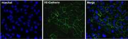
- Immunofluorescent analysis of VE-Cadherin (green) in Huvec cells. The cells were fixed with 4% paraformaldehyde in PBS for 15 minutes at room temperature, permeabilized with 0.1% Triton X-100 for 15 minutes, and blocked with 3% BSA for 30 minutes at room temperature. Cells were stained with a VE-Cadherin mouse monoclonal antibody (Product # MA1-198) at a concentration of 3 µg/mL in blocking buffer for 1 hour at room temperature, and then incubated with a Goat anti-Mouse IgG (H+L) Superclonal™ Secondary Antibody, Alexa Fluor® 488 conjugate (Product # A28175) at a dilution of 1:1000 for at least 30 minutes at a room temperature in the dark (green). Nuclei (blue) were stained with Hoechst 33342 (Product # 62249). Images were taken on a Thermo Scientific ToxInsight Instrument at 20X magnification.

Supportive validation
- Submitted by
- Invitrogen Antibodies (provider)
- Main image

- Experimental details
- Flow cytometry analysis of VE-Cadherin was done on Huvec cells. The cells were stained with a VE-Cadherin mouse monoclonal antibody (Product # MA1-198, red histogram) at a concentration of 1 µg/mL. After incubation of the primary antibody on ice for an hour, the cells were stained with a Goat anti-Mouse IgG (H+L) Secondary Antibody, Alexa Fluor Plus 680 conjugate (Product # A32734) at a dilution of 1:50 for at least 30 minutes on ice. A representative 10,000 cells were acquired for each sample. The black histogram represents unstained control cells and the blue histogram represents no-primary-antibody control.

Supportive validation
- Submitted by
- Invitrogen Antibodies (provider)
- Main image

- Experimental details
- NULL

- Submitted by
- Invitrogen Antibodies (provider)
- Main image

- Experimental details
- NULL
- Submitted by
- Invitrogen Antibodies (provider)
- Main image

- Experimental details
- NULL
