 Explore
 Validate
 Learn
Learn Western blot
Antibody data
- Antibody Data
- Antigen structure
- References [5]
- Comments [0]
- Validations
- Western blot [2]
- Immunocytochemistry [2]
- Other assay [4]
Submit
Validation data
Reference
Comment
Report error
- Product number
- PA5-19612 - Provider product page

- Provider
- Invitrogen Antibodies
- Product name
- VE-cadherin Polyclonal Antibody
- Antibody type
- Polyclonal
- Antigen
- Synthetic peptide
- Description
- Heat mediated antigen retrieval with Tris/EDTA buffer, pH 9.0, recommended prior to tissue staining. This antibody is predicted to react with cow and pig based on sequence homology. Store antibody at 4ºC for 1-2 weeks. For long-term storage, store at -20ºC.
- Reactivity
- Human, Mouse
- Host
- Rabbit
- Isotype
- IgG
- Vial size
- 100 µg
- Concentration
- 1.0 mg/mL
- Storage
- -20°C or -80°C if preferred
Submitted references Probing Endothelial Cell Mechanics Through Connexin 43 Disruption.
Post-ischaemic administration of the murine Canakinumab-surrogate antibody improves outcome in experimental stroke.
Inhibition of MicroRNA-155 Supports Endothelial Tight Junction Integrity Following Oxygen-Glucose Deprivation.
Sera From Children After Cardiopulmonary Bypass Reduces Permeability of Capillary Endothelial Cell Barriers.
HIF-1alpha Deficiency Attenuates the Cardiomyogenesis of Mouse Embryonic Stem Cells.
Islam MM, Steward RL Jr
Experimental mechanics 2019 Mar;59(3):327-336
Experimental mechanics 2019 Mar;59(3):327-336
Post-ischaemic administration of the murine Canakinumab-surrogate antibody improves outcome in experimental stroke.
Liberale L, Diaz-Cañestro C, Bonetti NR, Paneni F, Akhmedov A, Beer JH, Montecucco F, Lüscher TF, Camici GG
European heart journal 2018 Oct 7;39(38):3511-3517
European heart journal 2018 Oct 7;39(38):3511-3517
Inhibition of MicroRNA-155 Supports Endothelial Tight Junction Integrity Following Oxygen-Glucose Deprivation.
Pena-Philippides JC, Gardiner AS, Caballero-Garrido E, Pan R, Zhu Y, Roitbak T
Journal of the American Heart Association 2018 Jun 26;7(13)
Journal of the American Heart Association 2018 Jun 26;7(13)
Sera From Children After Cardiopulmonary Bypass Reduces Permeability of Capillary Endothelial Cell Barriers.
Pierce RW, Zahr RA, Kandil S, Faustino EVS, Pober JS
Pediatric critical care medicine : a journal of the Society of Critical Care Medicine and the World Federation of Pediatric Intensive and Critical Care Societies 2018 Jul;19(7):609-618
Pediatric critical care medicine : a journal of the Society of Critical Care Medicine and the World Federation of Pediatric Intensive and Critical Care Societies 2018 Jul;19(7):609-618
HIF-1alpha Deficiency Attenuates the Cardiomyogenesis of Mouse Embryonic Stem Cells.
Kudová J, Procházková J, Vašiček O, Perečko T, Sedláčková M, Pešl M, Pacherník J, Kubala L
PloS one 2016;11(6):e0158358
PloS one 2016;11(6):e0158358
No comments: Submit comment
Supportive validation
- Submitted by
- Invitrogen Antibodies (provider)
- Main image

- Experimental details
- Western blot analysis of Human Kidney Tissue Lysate using Product # PA5-19612, VE Cadherin primary antibody at a dilution of 1 µg/mL. Blot treated with a secondary IR Dye680-conjugated Goat polyclonal anti-Rabbit antibody was used at a dilution of 1:10000.

- Submitted by
- Invitrogen Antibodies (provider)
- Main image

- Experimental details
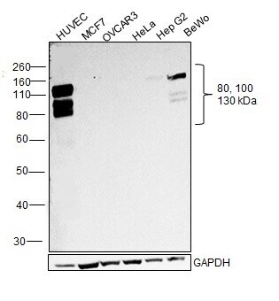
- Western blot was performed using Anti-VE-cadherin Polyclonal Antibody (Product # PA5-19612) and ~80,100,130 kDa bands corresponding to Cadherin-5 and its glycosylated forms, were observed across high expressing cell lines (HUVEC and BeWo) tested, and not in low expressing cell lines (MCF7, OVCAR3, HeLa and Hep G2). Whole extracts (Scraped) (30 µg lysate) of HUVEC (Lane 1), MCF7 (Lane 2), OVCAR3 (Lane 3), HeLa (Lane 4), Hep G2 (Lane 5) and BeWo (Lane 6) were electrophoresed using NuPAGE™ 10% Bis-Tris Protein Gel (Product # NP0302BOX). Resolved proteins were then transferred onto a Nitrocellulose membrane (Product # IB23001) by iBlot® 2 Dry Blotting System (Product # IB21001). The blot was probed with the primary antibody (1 µg/mL) and detected by chemiluminescence with Goat anti-Rabbit IgG (H+L) Superclonal™ Recombinant Secondary Antibody, HRP (Product # A27036,1:4000 dilution) using the iBright FL 1000 (Product # A32752). Chemiluminescent detection was performed using Novex® ECL Chemiluminescent Substrate Reagent Kit (Product # WP20005).
Supportive validation
- Submitted by
- Invitrogen Antibodies (provider)
- Main image

- Experimental details
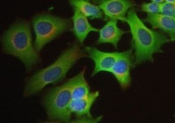
- Immunofluorescent staining of MCF-7 cells using Product # PA5-19612, anti-VE Cadherin antibody. The cells were fixed with methanol (100%) for 5 minutes, permabilised with TBS-T (20mins), BSA (1%), normal goat serum (10%) and glycine (0.3 M) in 0.1% PBS-Tween for 1 hour and exposed to the primary antibody at a concentration of 5 µg/mL overnight at 4C. The secondary antibody was a 448 fluorescence conjugated Goat anti-rabbit IgG (green) at a dilution of 1:1000. A WGA- 594 fluorescent conjugated stain was used to label plasma membranes (red) and the nuclei stain was DAPI (blue).

- Submitted by
- Invitrogen Antibodies (provider)
- Main image
- Experimental details
- Immunofluorescent staining of MCF-7 cells using Product # PA5-19612, anti-VE Cadherin antibody. The cells were fixed with methanol (100%) for 5 minutes, permabilised with TBS-T (20mins), BSA (1%), normal goat serum (10%) and glycine (0.3 M) in 0.1% PBS-Tween for 1 hour and exposed to the primary antibody at a concentration of 5 µg/mL overnight at 4C. The secondary antibody was a 448 fluorescence conjugated Goat anti-rabbit IgG (green) at a dilution of 1:1000. A WGA- 594 fluorescent conjugated stain was used to label plasma membranes (red) and the nuclei stain was DAPI (blue).

Supportive validation
- Submitted by
- Invitrogen Antibodies (provider)
- Main image

- Experimental details
- NULL

- Submitted by
- Invitrogen Antibodies (provider)
- Main image

- Experimental details
- NULL

- Submitted by
- Invitrogen Antibodies (provider)
- Main image

- Experimental details
- NULL
- Submitted by
- Invitrogen Antibodies (provider)
- Main image
- Experimental details
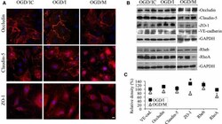
- Figure 4 Effect of microRNA-155 (miR-155) inhibition and overexpression on the endothelial cell junction proteins. Immunofluorescence microscopy (A) and Western blot analysis (B) were performed to detect the cellular distribution and expression levels of cell junction proteins in oxygen-glucose deprivation (OGD)/control inhibitor (OGD/IC), specific miR-155 inhibitor (OGD/I), and mimic (OGD/M) cells. A, Immunofluorescence staining for occludin, claudin-5, and zonula occludens protein-1 (ZO-1; red). Confocal microscopy images were acquired using a Zeiss LSM 800 confocal microscope. Bar: 10 mum. B, Representative immunoblots demonstrate the levels of occludin, claudin-5, ZO -1, and vascular endothelial (VE)-cadherin. In addition to cell junction proteins, Western blots were performed to assess expression of miR-155 direct target Rheb and RhoA bands. C, Graph demonstrates quantification of Western blot data obtained in OGD /I (black squares) and OGD /M (grey triangles) samples. Optical density of the protein bands was measured using ImageJ software, normalized to GAPDH density in every sample, and expressed as a percentage of the optical density calculated in the appropriate control OGD / IC and mimic control ( OGD / MC ) samples, respectively. n=4 (for OGD / IC and OGD / MC groups) and 6 (for OGD /I and OGD /M groups) independent experiments. Mann-Whitney (Wilcoxon) test was used to compare the relative protein levels between OGD /I and OGD / IC groups ( P =0.029). Error bars:
