Explore
 Validate
 Learn
Learn Western blot
Antibody data
- Antibody Data
- Antigen structure
- References [3]
- Comments [0]
- Validations
- Western blot [1]
- Immunocytochemistry [2]
Submit
Validation data
Reference
Comment
Report error
- Product number
- PA5-17401 - Provider product page

- Provider
- Invitrogen Antibodies
- Product name
- VE-cadherin Polyclonal Antibody
- Antibody type
- Polyclonal
- Antigen
- Synthetic peptide
- Description
- It is not recommended to aliquot this antibody.
- Concentration
- 22 µg/mL
Submitted references FTY720 Protects Against Ischemia-Reperfusion Injury by Preventing the Redistribution of Tight Junction Proteins and Decreases Inflammation in the Subacute Phase in an Experimental Stroke Model.
Tracking adiponectin biodistribution via fluorescence molecular tomography indicates increased vascular permeability after streptozotocin-induced diabetes.
Extracellular S100A4 affects endothelial cell integrity and stimulates transmigration of A375 melanoma cells.
Wang Z, Higashikawa K, Yasui H, Kuge Y, Ohno Y, Kihara A, Midori YA, Houkin K, Kawabori M
Translational stroke research 2020 Oct;11(5):1103-1116
Translational stroke research 2020 Oct;11(5):1103-1116
Tracking adiponectin biodistribution via fluorescence molecular tomography indicates increased vascular permeability after streptozotocin-induced diabetes.
Yoon N, Dadson K, Dang T, Chu T, Noskovicova N, Hinz B, Raignault A, Thorin E, Kim S, Jeon JS, Jonkman J, McKee TD, Grant J, Peterson JD, Kelly SP, Sweeney G
American journal of physiology. Endocrinology and metabolism 2019 Nov 1;317(5):E760-E772
American journal of physiology. Endocrinology and metabolism 2019 Nov 1;317(5):E760-E772
Extracellular S100A4 affects endothelial cell integrity and stimulates transmigration of A375 melanoma cells.
Herwig N, Belter B, Pietzsch J
Biochemical and biophysical research communications 2016 Sep 2;477(4):963-969
Biochemical and biophysical research communications 2016 Sep 2;477(4):963-969
No comments: Submit comment
Supportive validation
- Submitted by
- Invitrogen Antibodies (provider)
- Main image

- Experimental details
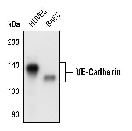
- Western blot analysis of VE-Cadherin in extracts from HUVEC and BAEC cells using VE-Cadherin polyclonal antibody (Product # PA5-17401).
Supportive validation
- Submitted by
- Invitrogen Antibodies (provider)
- Main image
- Experimental details
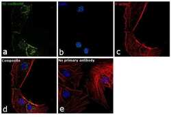
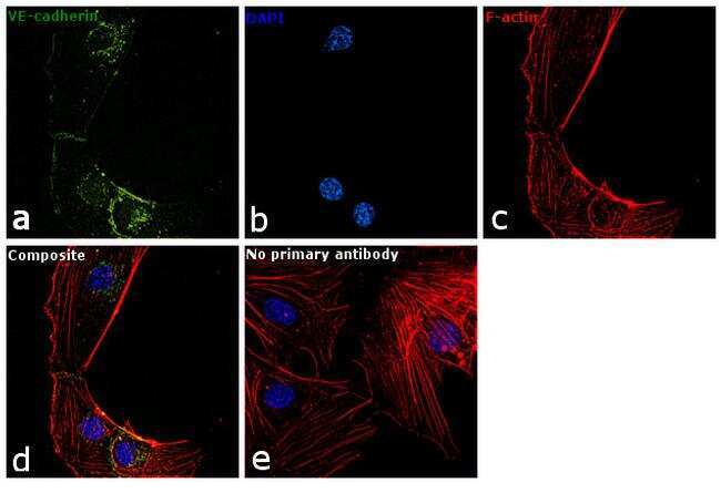
- Immunofluorescence analysis of VE-cadherin was performed using 70% confluent log phase HUVEC cells. The cells were fixed with 4% paraformaldehyde for 10 minutes, permeabilized with 0.1% Triton™ X-100 for 15 minutes, and blocked with 1% BSA for 1 hour at room temperature. The cells were labeled with VE-cadherin Polyclonal Antibody (Product # PA5-17401) at 1:100 dilution in 0.1% BSA, incubated at 4 degree Celsius overnight and then labeled with Goat anti-Rabbit IgG (H+L) Superclonal™ Secondary Antibody, Alexa Fluor® 488 conjugate (Product # A27034) at a dilution of 1:2000 for 45 minutes at room temperature (Panel a: green). Nuclei (Panel b: blue) were stained with SlowFade® Gold Antifade Mountant with DAPI (Product # S36938). F-actin (Panel c: red) was stained with Rhodamine Phalloidin (Product # R415, 1:300). Panel d represents the merged image showing cytoplasmic punctas and plasma membrane localization. Panel e represents control cells with no primary antibody to assess background. The images were captured at 60X magnification.
- Submitted by
- Invitrogen Antibodies (provider)
- Main image
- Experimental details
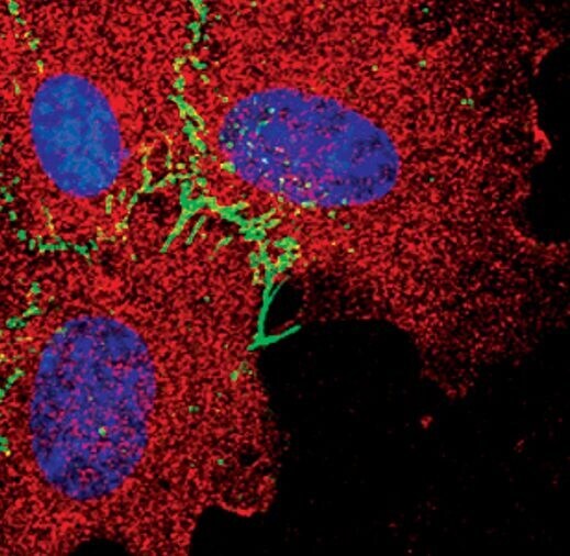
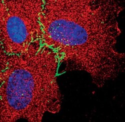
- Immunofluorescent analysis of VE-Cadherin in HUVE cells using a VE-Cadherin polyclonal antibody (Product # PA5-17401) (green) and a MEK1/2 monoclonal antibody (red). DNA is labeled using a fluorescent blue dye.