Explore
 Validate
 Learn
Learn Western blot
 Flow cytometry
Antibody data
- Antibody Data
- Antigen structure
- References [7]
- Comments [0]
- Validations
- Western blot [1]
- Immunocytochemistry [2]
Submit
Validation data
Reference
Comment
Report error
- Product number
- AF938 - Provider product page

- Provider
- R&D Systems
- Product name
- Human VE-Cadherin Antibody
- Antibody type
- Polyclonal
- Description
- Antigen Affinity-purified. Detects human VE-Cadherin in direct ELISAs and Western blots. In direct ELISAs, approximately 10% cross-reactivity with recombinant mouse VE-Cadherin is observed and less than 5% cross-reactivity with recombinant human E-Cadherin is observed.
- Reactivity
- Human
- Host
- Goat
- Conjugate
- Unconjugated
- Antigen sequence
P33151- Isotype
- IgG
- Vial size
- 100 ug
- Concentration
- LYOPH
- Storage
- Use a manual defrost freezer and avoid repeated freeze-thaw cycles. 12 months from date of receipt, -20 to -70 °C as supplied. 1 month, 2 to 8 °C under sterile conditions after reconstitution. 6 months, -20 to -70 °C under sterile conditions after reconstitution.
Submitted references aPKC controls endothelial growth by modulating c-Myc via FoxO1 DNA-binding ability.
Mechanically activated ion channel PIEZO1 is required for lymphatic valve formation.
Vascular CXCR4 Expression Promotes Vessel Sprouting and Sensitivity to Sorafenib Treatment in Hepatocellular Carcinoma.
DeepCAGE transcriptomics identify HOXD10 as a transcription factor regulating lymphatic endothelial responses to VEGF-C.
MicroRNA miR-27b rescues bone marrow-derived angiogenic cell function and accelerates wound healing in type 2 diabetes mellitus.
A novel serum-free monolayer culture for orderly hematopoietic differentiation of human pluripotent cells via mesodermal progenitors.
Signaling hierarchy regulating human endothelial cell development.
Riddell M, Nakayama A, Hikita T, Mirzapourshafiyi F, Kawamura T, Pasha A, Li M, Masuzawa M, Looso M, Steinbacher T, Ebnet K, Potente M, Hirose T, Ohno S, Fleming I, Gattenlöhner S, Aung PP, Phung T, Yamasaki O, Yanagi T, Umemura H, Nakayama M
Nature communications 2018 Dec 17;9(1):5357
Nature communications 2018 Dec 17;9(1):5357
Mechanically activated ion channel PIEZO1 is required for lymphatic valve formation.
Nonomura K, Lukacs V, Sweet DT, Goddard LM, Kanie A, Whitwam T, Ranade SS, Fujimori T, Kahn ML, Patapoutian A
Proceedings of the National Academy of Sciences of the United States of America 2018 Dec 11;115(50):12817-12822
Proceedings of the National Academy of Sciences of the United States of America 2018 Dec 11;115(50):12817-12822
Vascular CXCR4 Expression Promotes Vessel Sprouting and Sensitivity to Sorafenib Treatment in Hepatocellular Carcinoma.
Xu J, Liang J, Meng YM, Yan J, Yu XJ, Liu CQ, Xu L, Zhuang SM, Zheng L
Clinical cancer research : an official journal of the American Association for Cancer Research 2017 Aug 1;23(15):4482-4492
Clinical cancer research : an official journal of the American Association for Cancer Research 2017 Aug 1;23(15):4482-4492
DeepCAGE transcriptomics identify HOXD10 as a transcription factor regulating lymphatic endothelial responses to VEGF-C.
Klein S, Dieterich LC, Mathelier A, Chong C, Sliwa-Primorac A, Hong YK, Shin JW, Lizio M, Itoh M, Kawaji H, Lassmann T, Daub CO, Arner E, FANTOM consortium, Carninci P, Hayashizaki Y, Forrest AR, Wasserman WW, Detmar M
Journal of cell science 2016 Jul 1;129(13):2573-85
Journal of cell science 2016 Jul 1;129(13):2573-85
MicroRNA miR-27b rescues bone marrow-derived angiogenic cell function and accelerates wound healing in type 2 diabetes mellitus.
Wang JM, Tao J, Chen DD, Cai JJ, Irani K, Wang Q, Yuan H, Chen AF
Arteriosclerosis, thrombosis, and vascular biology 2014 Jan;34(1):99-109
Arteriosclerosis, thrombosis, and vascular biology 2014 Jan;34(1):99-109
A novel serum-free monolayer culture for orderly hematopoietic differentiation of human pluripotent cells via mesodermal progenitors.
Niwa A, Heike T, Umeda K, Oshima K, Kato I, Sakai H, Suemori H, Nakahata T, Saito MK
PloS one 2011;6(7):e22261
PloS one 2011;6(7):e22261
Signaling hierarchy regulating human endothelial cell development.
Kelly MA, Hirschi KK
Arteriosclerosis, thrombosis, and vascular biology 2009 May;29(5):718-24
Arteriosclerosis, thrombosis, and vascular biology 2009 May;29(5):718-24
No comments: Submit comment
Supportive validation
- Submitted by
- R&D Systems (provider)
- Main image

- Experimental details
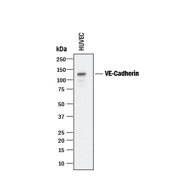
- Detection of Human VE-Cadherin by Western Blot. Western blot shows lysate of HUVEC human umbilical vein endothelial cells. PVDF membrane was probed with 0.25 µg/mL of Goat Anti-Human VE-Cadherin Antigen Affinity-purified Polyclonal Antibody (Catalog # AF938) followed by HRP-conjugated Anti-Goat IgG Secondary Antibody (Catalog # HAF017). A specific band was detected for VE-Cadherin at approximately 125 kDa (as indicated). This experiment was conducted under reducing conditions and using Immunoblot Buffer Group 1.
Supportive validation
- Submitted by
- R&D Systems (provider)
- Main image
- Experimental details
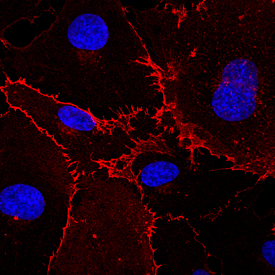
- VE-Cadherin in HUVEC Cells. VE-Cadherin was detected in immersion fixed HUVEC human umbilical vein endothelial cells using Goat Anti-Human VE-Cadherin Antigen Affinity-purified Polyclonal Antibody (Catalog # AF938) at 10 µg/mL for 3 hours at room temperature. Cells were stained using the NorthernLights™ 557-conjugated Anti-Goat IgG Secondary Antibody (red; Catalog # NL001) and counterstained with DAPI (blue). Specific staining was localized to the plasma membrane. View our protocol for Fluorescent ICC Staining of Cells on Coverslips.
- Submitted by
- R&D Systems (provider)
- Main image

- Experimental details
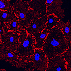
- VE-Cadherin in HUVEC Cells. VE-Cadherin was detected in immersion fixed HUVEC human umbilical vein endothelial cells using Goat Anti-Human VE-Cadherin Antigen Affinity-purified Polyclonal Antibody (Catalog # AF938) at 10 µg/mL for 3 hours at room temperature. Cells were stained using the NorthernLights™ 557-conjugated Anti-Goat IgG Secondary Antibody (red; Catalog # NL001) and counterstained with DAPI (blue). Specific staining was localized to the plasma membrane. View our protocol for Fluorescent ICC Staining of Cells on Coverslips.