Explore
 Validate
 Learn
Learn Western blot
 Flow cytometry
Antibody data
- Antibody Data
- Antigen structure
- References [8]
- Comments [0]
- Validations
- Western blot [1]
- Immunocytochemistry [1]
Submit
Validation data
Reference
Comment
Report error
- Product number
- MAB9381 - Provider product page

- Provider
- R&D Systems
- Product name
- Human VE-Cadherin Antibody
- Antibody type
- Monoclonal
- Description
- Protein A or G purified from hybridoma culture supernatant. Detects human VE-Cadherin in Western blots. In Western blots, 25% cross-reactivity with recombinant mouse VE-Cadherin and no cross-reactivity with recombinant human (rh) Cadherin-17 or rhP-Cadherin is observed.
- Reactivity
- Human
- Host
- Mouse
- Conjugate
- Unconjugated
- Antigen sequence
P33151- Isotype
- IgG
- Antibody clone number
- 123413
- Vial size
- 100 ug
- Concentration
- LYOPH
- Storage
- Use a manual defrost freezer and avoid repeated freeze-thaw cycles. 12 months from date of receipt, -20 to -70 °C as supplied. 1 month, 2 to 8 °C under sterile conditions after reconstitution. 6 months, -20 to -70 °C under sterile conditions after reconstitution.
Submitted references Functional 3D Human Liver Bud Assembled from MSC-Derived Multiple Liver Cell Lineages.
Age-Related Changes in HAPLN1 Increase Lymphatic Permeability and Affect Routes of Melanoma Metastasis.
Human pre-valvular endocardial cells derived from pluripotent stem cells recapitulate cardiac pathophysiological valvulogenesis.
The Superantigen Toxic Shock Syndrome Toxin 1 Alters Human Aortic Endothelial Cell Function.
Synergic effects of VEGF-A and SDF-1 on the angiogenic properties of endothelial progenitor cells.
A Three-Dimensional Cell Culture System To Model RNA Virus Infections at the Blood-Brain Barrier.
Mesp1 coordinately regulates cardiovascular fate restriction and epithelial-mesenchymal transition in differentiating ESCs.
Cannabidiol attenuates high glucose-induced endothelial cell inflammatory response and barrier disruption.
Li J, Xing F, Chen F, He L, So KF, Liu Y, Xiao J
Cell transplantation 2019 May;28(5):510-521
Cell transplantation 2019 May;28(5):510-521
Age-Related Changes in HAPLN1 Increase Lymphatic Permeability and Affect Routes of Melanoma Metastasis.
Ecker BL, Kaur A, Douglass SM, Webster MR, Almeida FV, Marino GE, Sinnamon AJ, Neuwirth MG, Alicea GM, Ndoye A, Fane M, Xu X, Sim MS, Deutsch GB, Faries MB, Karakousis GC, Weeraratna AT
Cancer discovery 2019 Jan;9(1):82-95
Cancer discovery 2019 Jan;9(1):82-95
Human pre-valvular endocardial cells derived from pluripotent stem cells recapitulate cardiac pathophysiological valvulogenesis.
Neri T, Hiriart E, van Vliet PP, Faure E, Norris RA, Farhat B, Jagla B, Lefrancois J, Sugi Y, Moore-Morris T, Zaffran S, Faustino RS, Zambon AC, Desvignes JP, Salgado D, Levine RA, de la Pompa JL, Terzic A, Evans SM, Markwald R, Pucéat M
Nature communications 2019 Apr 26;10(1):1929
Nature communications 2019 Apr 26;10(1):1929
The Superantigen Toxic Shock Syndrome Toxin 1 Alters Human Aortic Endothelial Cell Function.
Kulhankova K, Kinney KJ, Stach JM, Gourronc FA, Grumbach IM, Klingelhutz AJ, Salgado-Pabón W
Infection and immunity 2018 Mar;86(3)
Infection and immunity 2018 Mar;86(3)
Synergic effects of VEGF-A and SDF-1 on the angiogenic properties of endothelial progenitor cells.
Odent Grigorescu G, Rosca AM, Preda MB, Tutuianu R, Simionescu M, Burlacu A
Journal of tissue engineering and regenerative medicine 2017 Nov;11(11):3241-3252
Journal of tissue engineering and regenerative medicine 2017 Nov;11(11):3241-3252
A Three-Dimensional Cell Culture System To Model RNA Virus Infections at the Blood-Brain Barrier.
Bramley JC, Drummond CG, Lennemann NJ, Good CA, Kim KS, Coyne CB
mSphere 2017 May-Jun;2(3)
mSphere 2017 May-Jun;2(3)
Mesp1 coordinately regulates cardiovascular fate restriction and epithelial-mesenchymal transition in differentiating ESCs.
Lindsley RC, Gill JG, Murphy TL, Langer EM, Cai M, Mashayekhi M, Wang W, Niwa N, Nerbonne JM, Kyba M, Murphy KM
Cell stem cell 2008 Jul 3;3(1):55-68
Cell stem cell 2008 Jul 3;3(1):55-68
Cannabidiol attenuates high glucose-induced endothelial cell inflammatory response and barrier disruption.
Rajesh M, Mukhopadhyay P, Bátkai S, Haskó G, Liaudet L, Drel VR, Obrosova IG, Pacher P
American journal of physiology. Heart and circulatory physiology 2007 Jul;293(1):H610-9
American journal of physiology. Heart and circulatory physiology 2007 Jul;293(1):H610-9
No comments: Submit comment
Supportive validation
- Submitted by
- R&D Systems (provider)
- Main image
- Experimental details
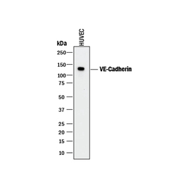
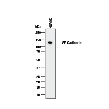
- Detection of Human VE-Cadherin by Western Blot. Western blot shows lysate of HUVEC human umbilical vein endothelial cells. PVDF membrane was probed with 1 µg/mL of Mouse Anti-Human VE-Cadherin Monoclonal Antibody (Catalog # MAB9381) followed by HRP-conjugated Anti-Mouse IgG Secondary Antibody (Catalog # HAF018). A specific band was detected for VE-Cadherin at approximately 125 kDa (as indicated). This experiment was conducted under reducing conditions and using Immunoblot Buffer Group 1.
Supportive validation
- Submitted by
- R&D Systems (provider)
- Main image

- Experimental details
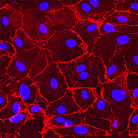
- VE-Cadherin in HUVEC Cells. VE-Cadherin was detected in immersion fixed HUVEC cells using Mouse Anti-Human VE-Cadherin Monoclonal Antibody (Catalog # MAB9381) at 0.5 µg/mL for 3 hours at room temperature. Cells were stained using the NorthernLights™ 557-conjugated Anti-Mouse IgG Secondary Antibody (red; Catalog # NL007) and counterstained with DAPI (blue). Specific staining was localized to plasma membrane. View our protocol for Fluorescent ICC Staining of Cells on Coverslips.