Explore
 Validate
 Learn
Learn Flow cytometry
Antibody data
- Antibody Data
- Antigen structure
- References [1]
- Comments [0]
- Validations
- Flow cytometry [1]
- Other assay [1]
Submit
Validation data
Reference
Comment
Report error
- Product number
- 12-1629-42 - Provider product page

- Provider
- Invitrogen Antibodies
- Product name
- CD162 (PSGL-1) Monoclonal Antibody (FLEG), PE, eBioscience™
- Antibody type
- Monoclonal
- Antigen
- Other
- Description
- Description: This FLEG monoclonal antibody recognizes human CD162, which is also known as P-selectin glyocoprotein ligand-1 (PSGL-1). This 120-kDa protein exists as a homodimer on the surface of monocytes, neutrophils, granulocytes, peripheral T cells, some B cells, and a subset of CD34+ hematopoietic progenitor cells (HPCs) in the bone marrow. CD162 binds CD62P (P-selectin), CD62E (E-selectin), and CD62L (L-selectin) to mediate leukocyte interactions with each other as well as with activated platelets and endothelium. Thus, CD162 plays a critical role in leukocyte adhesion and rolling. Furthermore, the binding of CD162 to P-selectin on HPCs has been demonstrated to inhibit hematopoiesis. Studies have also demonstrated CD162 as the receptor for enterovirus 71. Applications Reported: This FLEG antibody has been reported for use in flow cytometric analysis. Applications Tested: This FLEG antibody has been pre-titrated and tested by flow cytometric analysis of normal human peripheral blood cells. This can be used at 5 µL (0.06 µg) per test. A test is defined as the amount (µg) of antibody that will stain a cell sample in a final volume of 100 µL. Cell number should be determined empirically but can range from 10^5 to 10^8 cells/test. Excitation: 488-561 nm; Emission: 578 nm; Laser: Blue Laser, Green Laser, Yellow-Green Laser. Filtration: 0.2 µm post-manufacturing filtered.
- Reactivity
- Human
- Host
- Mouse
- Conjugate
- Yellow dye
- Isotype
- IgG
- Antibody clone number
- FLEG
- Vial size
- 100 Tests
- Concentration
- 5 µL/Test
- Storage
- 4° C, store in dark, DO NOT FREEZE!
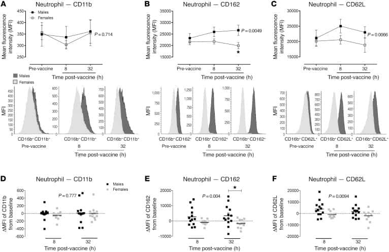
Submitted references Accelerated resolution of inflammation underlies sex differences in inflammatory responses in humans.
Rathod KS, Kapil V, Velmurugan S, Khambata RS, Siddique U, Khan S, Van Eijl S, Gee LC, Bansal J, Pitrola K, Shaw C, D'Acquisto F, Colas RA, Marelli-Berg F, Dalli J, Ahluwalia A
The Journal of clinical investigation 2017 Jan 3;127(1):169-182
The Journal of clinical investigation 2017 Jan 3;127(1):169-182
No comments: Submit comment
Supportive validation
- Submitted by
- Invitrogen Antibodies (provider)
- Main image
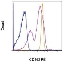
- Experimental details
- Staining of normal human peripheral blood cells with Mouse IgG2a K Isotype Control PE (Product # 12-4724-81) (blue histogram) or Anti-Human CD162 (PSGL-1) PE (purple histogram). Cells in the lymphocyte gate were used for analysis. The orange histogram shows staining of Anti-Human CD162 (PSGL-1) PE on granulocytes.
- Conjugate
- Yellow dye

Supportive validation
- Submitted by
- Invitrogen Antibodies (provider)
- Main image
- Experimental details
- NULL
- Conjugate
- Yellow dye