Explore
 Validate
 Learn
Learn Western blot
Antibody data
- Antibody Data
- Antigen structure
- References [0]
- Comments [0]
- Validations
- Western blot [1]
- Immunocytochemistry [1]
- Immunohistochemistry [1]
- Flow cytometry [1]
Submit
Validation data
Reference
Comment
Report error
- Product number
- MAB9961 - Provider product page

- Provider
- R&D Systems
- Product name
- Human PSGL-1/CD162 Antibody
- Antibody type
- Monoclonal
- Description
- Protein A or G purified from hybridoma culture supernatant. Detects human PSGL-1/CD162 in direct ELISAs and Western blots.
- Reactivity
- Human
- Host
- Mouse
- Conjugate
- Unconjugated
- Antigen sequence
NP_002997- Isotype
- IgG
- Antibody clone number
- 688101
- Vial size
- 100 ug
- Concentration
- LYOPH
- Storage
- Use a manual defrost freezer and avoid repeated freeze-thaw cycles. 12 months from date of receipt, -20 to -70 °C as supplied. 1 month, 2 to 8 °C under sterile conditions after reconstitution. 6 months, -20 to -70 °C under sterile conditions after reconstitution.
No comments: Submit comment
Supportive validation
- Submitted by
- R&D Systems (provider)
- Main image
- Experimental details
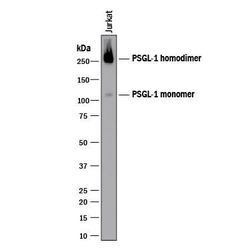
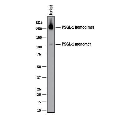
- Detection of Human PSGL-1/CD162 by Western Blot. Western blot shows lysates of Jurkat human acute T cell leukemia cell line. PVDF membrane was probed with 2 µg/mL of Mouse Anti-Human PSGL-1/CD162 Monoclonal Antibody (Catalog # MAB9961) followed by HRP-conjugated Anti-Mouse IgG Secondary Antibody (Catalog # HAF018). Specific bands were detected for PSGL1/ CD162 homodimer at approximately 250 kDa and PSGL1/ CD162 monomer at approximately 110 kDa (as indicated). This experiment was conducted under reducing conditions and using Immunoblot Buffer Group 1.
Supportive validation
- Submitted by
- R&D Systems (provider)
- Main image
- Experimental details
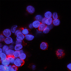
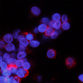
- PSGL-1/CD162 in Human PBMCs. PSGL-1/CD162 was detected in immersion fixed human peripheral blood mononuclear cells (PBMCs) using Mouse Anti-Human PSGL-1/CD162 Monoclonal Antibody (Catalog # MAB9961) at 8 µg/mL for 3 hours at room temperature. Cells were stained using the NorthernLights™ 557-conjugated Anti-Mouse IgG Secondary Antibody (red; Catalog # NL007) and counterstained with DAPI (blue). Specific staining was localized to cytoplasm. View our protocol for Fluorescent ICC Staining of Non-adherent Cells.
Supportive validation
- Submitted by
- R&D Systems (provider)
- Main image
- Experimental details
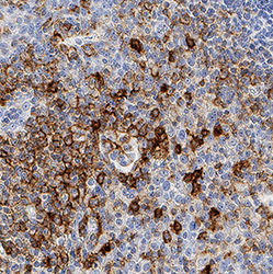
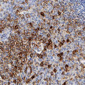
- PSGL-1/CD162 in Human Tonsil. PSGL-1/CD162 was detected in immersion fixed paraffin-embedded sections of human tonsil using Mouse Anti-Human PSGL-1/CD162 Monoclonal Antibody (Catalog # MAB9961) at 5 µg/mL for 1 hour at room temperature followed by incubation with the Anti-Mouse IgG VisUCyte™ HRP Polymer Antibody (Catalog # VC001). Tissue was stained using DAB (brown) and counterstained with hematoxylin (blue). Specific staining was localized to cytoplasm. View our protocol for IHC Staining with VisUCyte HRP Polymer Detection Reagents.
Supportive validation
- Submitted by
- R&D Systems (provider)
- Main image
- Experimental details
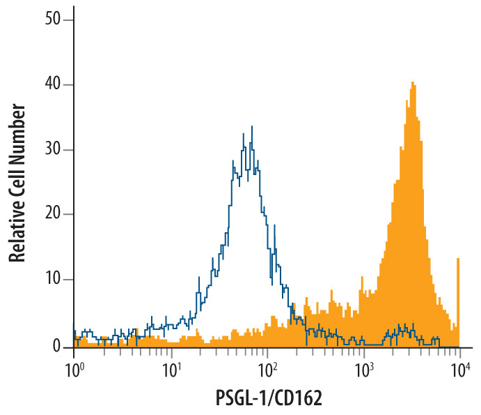
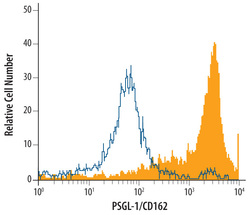
- Detection of PSGL-1 in Human Peripheral Blood Monocytes by Flow Cytometry. Human peripheral blood monocytes were stained with Mouse Anti-Human PSGL-1/CD162 Monoclonal Antibody (Catalog # MAB9961, filled histogram) or isotype control antibody (Catalog # MAB003, open histogram), followed by Allophycocyanin-conjugated Anti-Mouse IgG Secondary Antibody (Catalog # F0101B).