 Explore
 Validate
 Learn
Learn Western blot
 ELISA
ELISAAntibody data
- Antibody Data
- Antigen structure
- References [1]
- Comments [0]
- Validations
- Western blot [2]
- Chromatin Immunoprecipitation [1]
Submit
Validation data
Reference
Comment
Report error
- Product number
- MA1-20354 - Provider product page

- Provider
- Invitrogen Antibodies
- Product name
- HDAC1 Monoclonal Antibody (HDAC1-21)
- Antibody type
- Monoclonal
- Antigen
- Synthetic peptide
- Description
- Light sensitivity: This tandem dye is sensitive to photo-induced oxidation. Please protect this vial and stained samples from light.
- Reactivity
- Human, Mouse
- Host
- Mouse
- Isotype
- IgG
- Antibody clone number
- HDAC1-21
- Vial size
- 100 µL
- Concentration
- 2.4 mg/mL
- Storage
- Store at 4°C short term. For long term storage, store at -20°C, avoiding freeze/thaw cycles.
Submitted references Adenylyl cyclase activating polypeptide reduces phosphorylation and toxicity of the polyglutamine-expanded androgen receptor in spinobulbar muscular atrophy.
Polanco MJ, Parodi S, Piol D, Stack C, Chivet M, Contestabile A, Miranda HC, Lievens PM, Espinoza S, Jochum T, Rocchi A, Grunseich C, Gainetdinov RR, Cato AC, Lieberman AP, La Spada AR, Sambataro F, Fischbeck KH, Gozes I, Pennuto M
Science translational medicine 2016 Dec 21;8(370):370ra181
Science translational medicine 2016 Dec 21;8(370):370ra181
No comments: Submit comment
Supportive validation
- Submitted by
- Invitrogen Antibodies (provider)
- Main image
- Experimental details
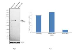
- Knockdown of HDAC1 was achieved by transfecting MCF7 cells with HDAC1 specific siRNAs (Silencer® select Product # s73). Western blot analysis (Fig. a) was performed using Modified whole cell extracts (1% SDS) from the HDAC1 knockdown cells (Lane 3), non-specific scrambled siRNA transfected cells (Lane 2) and untransfected cells (Lane 1). The blot was probed with Anti-HDAC1 Monoclonal Antibody (HDAC1-21) (Product # MA1-20354, 2µg/ml) and Goat anti-Mouse IgG (H+L) Superclonal™ Recombinant Secondary Antibody, HRP (Product # A28177, 1:4000 dilution) using the iBright FL 1000 (Product # A32752). Densitometric analysis of this Western Blot is shown in histogram (Fig. b). Decrease in signal upon siRNA mediated knock down confirms that antibody is specific to HDAC1..

- Submitted by
- Invitrogen Antibodies (provider)
- Main image

- Experimental details
- Western blot was performed using Anti-HDAC1 Polyclonal Antibody (Product # MA1-20354) and ~55kDa band corresponding to HDAC1 was observed in MCF-7, HeLa, HEK-293 and NIH/3T3 cells. Modified whole cell extracts (1% SDS) (30 ug lysate) of MCF-7 (Lane 1), HeLa (Lane 2), HEK-293 (Lane 3) and NIH/3T3 (Lane 4) were electrophoresed using NuPAGE® 4-12% Bis-Tris gel (Product # NP0322BOX). Resolved proteins were then transferred onto a nitrocellulose membrane (Product # IB23001) by iBlot® 2 Dry Blotting System (Product # IB21001).The blot was probed with the primary antibody (2µg/ml) and detected by chemiluminescence with Goat anti-Mouse IgG (H+L) Superclonal™ Recombinant Secondary Antibody, HRP (Product # A28177, 1:4000 dilution) using the iBright FL 1000 (Product # A32752). Chemiluminescent detection was performed using Novex® ECL Chemiluminescent Substrate Reagent Kit (Product # WP20005)..

Supportive validation
- Submitted by
- Invitrogen Antibodies (provider)
- Main image

- Experimental details
- Chromatin Immunoprecipitation (ChIP) assay of endogenous HDAC1 protein using Anti-HDAC1 Antibody: ChIP was performed using Anti-HDAC1 Mouse Monoclonal Antibody (Product # MA1-20354, 5 µg) on sheared chromatin from HeLa cells using the MAGnify ChIP System kit (Product # 49-2024). Normal Mouse IgG was used as a negative IP control. The purified DNA was analyzed by qPCR using primers binding to CDKN1A Intron 1 and SATA satellite repeats. Data is presented as fold enrichment of the antibody signal versus the negative control IgG using the comparative CT method.
