Explore
 Validate
 Learn
Learn Western blot
 Immunocytochemistry
Antibody data
- Antibody Data
- Antigen structure
- References [2]
- Comments [0]
- Validations
- Western blot [1]
- Immunocytochemistry [1]
- Immunohistochemistry [1]
Submit
Validation data
Reference
Comment
Report error
- Product number
- HPA029693 - Provider product page

- Provider
- Atlas Antibodies
- Proper citation
- Atlas Antibodies Cat#HPA029693, RRID:AB_10601659
- Product name
- Anti-HDAC1
- Antibody type
- Polyclonal
- Description
- Polyclonal Antibody against Human HDAC1, Gene description: histone deacetylase 1, Alternative Gene Names: GON-10, HD1, RPD3L1, Validated applications: WB, IHC, ICC, Uniprot ID: Q13547, Storage: Store at +4°C for short term storage. Long time storage is recommended at -20°C.
- Reactivity
- Human
- Host
- Rabbit
- Conjugate
- Unconjugated
- Isotype
- IgG
- Vial size
- 100 µl
- Concentration
- 0.2 mg/ml
- Storage
- Store at +4°C for short term storage. Long time storage is recommended at -20°C.
- Handling
- The antibody solution should be gently mixed before use.
Submitted references Aggregation and disaggregation features of the human proteome
Recruitment of Oct4 Protein to UV-Damaged Chromatin in Embryonic Stem Cells
Määttä T, Rettel M, Sridharan S, Helm D, Kurzawa N, Stein F, Savitski M
Molecular Systems Biology 2020;16(10)
Molecular Systems Biology 2020;16(10)
Recruitment of Oct4 Protein to UV-Damaged Chromatin in Embryonic Stem Cells
Cotterill S, Bártová E, Šustáčková G, Stixová L, Kozubek S, Legartová S, Foltánková V
PLoS ONE 2011;6(12):e27281
PLoS ONE 2011;6(12):e27281
No comments: Submit comment
Enhanced validation
- Submitted by
- Atlas Antibodies (provider)
- Enhanced method
- Genetic validation
- Main image
- Experimental details
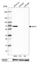
- Western blot analysis in RT-4 cells transfected with control siRNA, target specific siRNA probe #1 and #2, using Anti-HDAC1 antibody. Remaining relative intensity is presented. Loading control: Anti-GAPDH.
- Sample type
- Human
- Protocol
- Protocol

Supportive validation
- Submitted by
- Atlas Antibodies (provider)
- Main image
- Experimental details
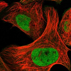
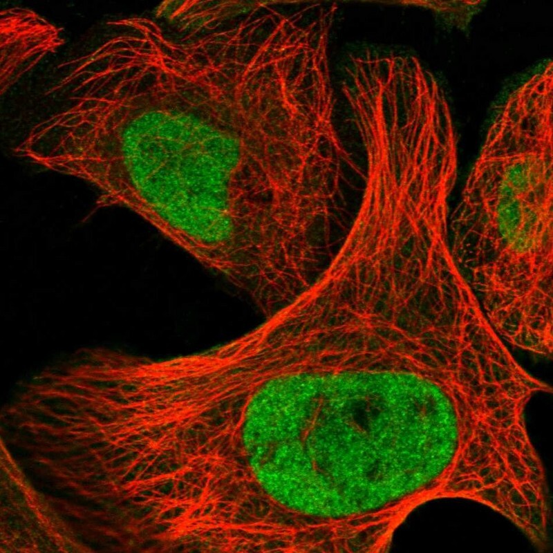
- Immunofluorescent staining of human cell line U-2 OS shows localization to nucleoplasm.
- Sample type
- Human
Supportive validation
- Submitted by
- Atlas Antibodies (provider)
- Enhanced method
- Orthogonal validation
- Main image
- Experimental details
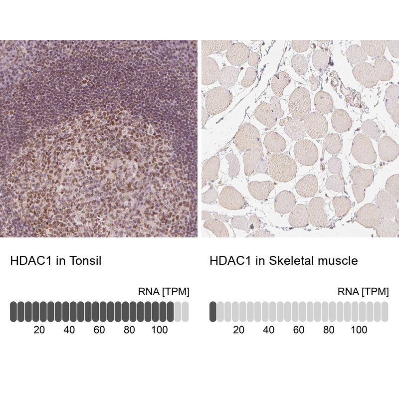
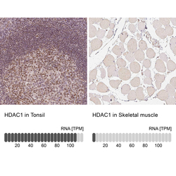
- Immunohistochemistry analysis in human tonsil and skeletal muscle tissues using HPA029693 antibody. Corresponding HDAC1 RNA-seq data are presented for the same tissues.
- Sample type
- Human
- Protocol
- Protocol