Explore
 Validate
 Learn
Learn Western blot
 Immunocytochemistry
Antibody data
- Antibody Data
- Antigen structure
- References [13]
- Comments [0]
- Validations
- Western blot [9]
- Immunocytochemistry [2]
- Immunoprecipitation [1]
- Immunohistochemistry [6]
- Chromatin Immunoprecipitation [1]
Submit
Validation data
Reference
Comment
Report error
- Product number
- GTX100513 - Provider product page

- Provider
- GeneTex
- Proper citation
- GeneTex Cat#GTX100513, RRID:AB_1240929
- Product name
- HDAC1 antibody
- Antibody type
- Polyclonal
- Reactivity
- Human, Mouse, Rat
- Host
- Rabbit
Submitted references The chromatin remodeler RSF1 controls centromeric histone modifications to coordinate chromosome segregation.
The anti-inflammatory effect of 10-oxo-trans-11-octadecenoic acid (KetoC) on RAW 264.7 cells stimulated with Porphyromonas gingivalis lipopolysaccharide.
NKILA lncRNA promotes tumor immune evasion by sensitizing T cells to activation-induced cell death.
Delaying histone deacetylase response to injury accelerates conversion into repair Schwann cells and nerve regeneration.
Inhibition of HDAC3- and HDAC6-Promoted Survivin Expression Plays an Important Role in SAHA-Induced Autophagy and Viability Reduction in Breast Cancer Cells.
Clinacanthus nutans Protects Cortical Neurons Against Hypoxia-Induced Toxicity by Downregulating HDAC1/6.
Immunohistochemical Characterization of Histone Deacetylase as a Potential Prognostic Marker and Therapeutic Target in Endometrial Stromal Sarcoma.
HDAC1/2-Dependent P0 Expression Maintains Paranodal and Nodal Integrity Independently of Myelin Stability through Interactions with Neurofascins.
cAMP-induced phosphorylation of 26S proteasomes on Rpn6/PSMD11 enhances their activity and the degradation of misfolded proteins.
A gene expression signature-based approach reveals the mechanisms of action of the Chinese herbal medicine berberine.
Nuclear proteomics with XRCC3 knockdown to reveal the development of doxorubicin-resistant uterine cancer.
Retinoic acid controls body axis extension by directly repressing Fgf8 transcription.
Role of autophagy in chemoresistance: regulation of the ATM-mediated DNA-damage signaling pathway through activation of DNA-PKcs and PARP-1.
Lee HS, Lin Z, Chae S, Yoo YS, Kim BG, Lee Y, Johnson JL, Kim YS, Cantley LC, Lee CW, Yu H, Cho H
Nature communications 2018 Sep 21;9(1):3848
Nature communications 2018 Sep 21;9(1):3848
The anti-inflammatory effect of 10-oxo-trans-11-octadecenoic acid (KetoC) on RAW 264.7 cells stimulated with Porphyromonas gingivalis lipopolysaccharide.
Sulijaya B, Takahashi N, Yamada M, Yokoji M, Sato K, Aoki-Nonaka Y, Nakajima T, Kishino S, Ogawa J, Yamazaki K
Journal of periodontal research 2018 Oct;53(5):777-784
Journal of periodontal research 2018 Oct;53(5):777-784
NKILA lncRNA promotes tumor immune evasion by sensitizing T cells to activation-induced cell death.
Huang D, Chen J, Yang L, Ouyang Q, Li J, Lao L, Zhao J, Liu J, Lu Y, Xing Y, Chen F, Su F, Yao H, Liu Q, Su S, Song E
Nature immunology 2018 Oct;19(10):1112-1125
Nature immunology 2018 Oct;19(10):1112-1125
Delaying histone deacetylase response to injury accelerates conversion into repair Schwann cells and nerve regeneration.
Brügger V, Duman M, Bochud M, Münger E, Heller M, Ruff S, Jacob C
Nature communications 2017 Jan 31;8:14272
Nature communications 2017 Jan 31;8:14272
Inhibition of HDAC3- and HDAC6-Promoted Survivin Expression Plays an Important Role in SAHA-Induced Autophagy and Viability Reduction in Breast Cancer Cells.
Lee JY, Kuo CW, Tsai SL, Cheng SM, Chen SH, Chan HH, Lin CH, Lin KY, Li CF, Kanwar JR, Leung EY, Cheung CC, Huang WJ, Wang YC, Cheung CH
Frontiers in pharmacology 2016;7:81
Frontiers in pharmacology 2016;7:81
Clinacanthus nutans Protects Cortical Neurons Against Hypoxia-Induced Toxicity by Downregulating HDAC1/6.
Tsai HD, Wu JS, Kao MH, Chen JJ, Sun GY, Ong WY, Lin TN
Neuromolecular medicine 2016 Sep;18(3):274-82
Neuromolecular medicine 2016 Sep;18(3):274-82
Immunohistochemical Characterization of Histone Deacetylase as a Potential Prognostic Marker and Therapeutic Target in Endometrial Stromal Sarcoma.
Baek MH, Park JY, Rhim CC, Park Y, Kim KR, Kim JH, Nam JH
Anticancer research 2016 May;36(5):2527-34
Anticancer research 2016 May;36(5):2527-34
HDAC1/2-Dependent P0 Expression Maintains Paranodal and Nodal Integrity Independently of Myelin Stability through Interactions with Neurofascins.
Brügger V, Engler S, Pereira JA, Ruff S, Horn M, Welzl H, Münger E, Vaquié A, Sidiropoulos PN, Egger B, Yotovski P, Filgueira L, Somandin C, Lühmann TC, D'Antonio M, Yamaguchi T, Matthias P, Suter U, Jacob C
PLoS biology 2015;13(9):e1002258
PLoS biology 2015;13(9):e1002258
cAMP-induced phosphorylation of 26S proteasomes on Rpn6/PSMD11 enhances their activity and the degradation of misfolded proteins.
Lokireddy S, Kukushkin NV, Goldberg AL
Proceedings of the National Academy of Sciences of the United States of America 2015 Dec 29;112(52):E7176-85
Proceedings of the National Academy of Sciences of the United States of America 2015 Dec 29;112(52):E7176-85
A gene expression signature-based approach reveals the mechanisms of action of the Chinese herbal medicine berberine.
Lee KH, Lo HL, Tang WC, Hsiao HH, Yang PM
Scientific reports 2014 Sep 17;4:6394
Scientific reports 2014 Sep 17;4:6394
Nuclear proteomics with XRCC3 knockdown to reveal the development of doxorubicin-resistant uterine cancer.
Chang JF, Lin ST, Hung E, Lu YL, Soon May EW, Lo YW, Chou HC, Chan HL
Toxicological sciences : an official journal of the Society of Toxicology 2014 Jun;139(2):396-406
Toxicological sciences : an official journal of the Society of Toxicology 2014 Jun;139(2):396-406
Retinoic acid controls body axis extension by directly repressing Fgf8 transcription.
Kumar S, Duester G
Development (Cambridge, England) 2014 Aug;141(15):2972-7
Development (Cambridge, England) 2014 Aug;141(15):2972-7
Role of autophagy in chemoresistance: regulation of the ATM-mediated DNA-damage signaling pathway through activation of DNA-PKcs and PARP-1.
Yoon JH, Ahn SG, Lee BH, Jung SH, Oh SH
Biochemical pharmacology 2012 Mar 15;83(6):747-57
Biochemical pharmacology 2012 Mar 15;83(6):747-57
No comments: Submit comment
Enhanced validation
Supportive validation
- Submitted by
- GeneTex (provider)
- Enhanced method
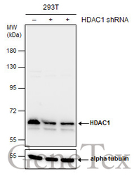
- Genetic validation
- Main image
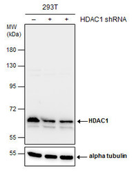
- Experimental details
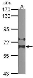
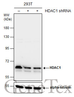
- Non-transfected (¡V) and transfected (+) 293T whole cell extracts (30 ?g) were separated by 7.5% SDS-PAGE, and the membrane was blotted with HDAC1 antibody (GTX100513) diluted at 1:4000. The HRP-conjugated anti-rabbit IgG antibody (GTX213110-01) was used to detect the primary antibody.
Supportive validation
- Submitted by
- GeneTex (provider)
- Main image
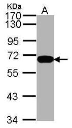
- Experimental details
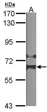
- Sample (30 ug of whole cell lysate) A: Hela 7.5% SDS PAGE HDAC1 antibody GTX100513 diluted at 1:1000
- Validation comment
- WB

- Submitted by
- GeneTex (provider)
- Main image
- Experimental details
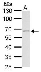
- Sample (30 ?g of whole cell lysate) A:NIH-3T37.5% SDS PAGE GTX100513 diluted at 1:1000 The HRP-conjugated anti-rabbit IgG antibody (GTX213110-01) was used to detect the primary antibody.
- Submitted by
- GeneTex (provider)
- Main image
- Experimental details
- HDAC1 antibody detects HDAC1 protein by western blot analysis.A. 30 ?g Rat2 whole cell lysate/extract10% SDS-PAGEHDAC1 antibody (GTX100513) dilution: 1:1000 The HRP-conjugated anti-rabbit IgG antibody (GTX213110-01) was used to detect the primary antibody.

- Submitted by
- GeneTex (provider)
- Main image
- Experimental details
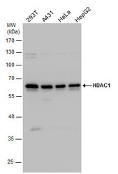
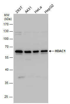
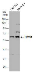
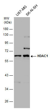
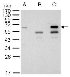
- HDAC1 antibody detects HDAC1 protein by western blot analysis. Various whole cell extracts (30 ?g) were separated by 10% SDS-PAGE, and the membrane was blotted with HDAC1 antibody (GTX100513) diluted by 1:1000. The HRP-conjugated anti-rabbit IgG antibody (GTX213110-01) was used to detect the primary antibody.
- Submitted by
- GeneTex (provider)
- Main image
- Experimental details
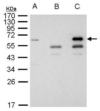
- Various whole cell extracts (30 ?g) were separated by 10% SDS-PAGE, and the membrane was blotted with HDAC1 antibody (GTX100513) diluted at 1:1000. The HRP-conjugated anti-rabbit IgG antibody (GTX213110-01) was used to detect the primary antibody.
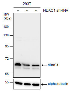
- Submitted by
- GeneTex (provider)
- Main image
- Experimental details
- Non-transfected (¡V) and transfected (+) 293T whole cell extracts (30 ?g) were separated by 7.5% SDS-PAGE, and the membrane was blotted with HDAC1 antibody (GTX100513) diluted at 1:4000. The HRP-conjugated anti-rabbit IgG antibody (GTX213110-01) was used to detect the primary antibody.
- Submitted by
- GeneTex (provider)
- Main image
- Experimental details
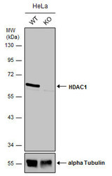
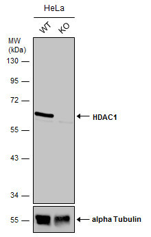
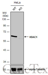
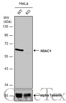
- Wild-type (WT) and HDAC1 knockout (KO) HeLa cell extracts (30 ?g) were separated by 10% SDS-PAGE, and the membrane was blotted with HDAC1 antibody (GTX100513) diluted at 1:500. The HRP-conjugated anti-rabbit IgG antibody (GTX213110-01) was used to detect the primary antibody.
- Submitted by
- GeneTex (provider)
- Main image
- Experimental details
- Wild-type (WT) and HDAC1 knockout (KO) HeLa cell extracts (30 ?g) were separated by 10% SDS-PAGE, and the membrane was blotted with HDAC1 antibody (GTX100513) diluted at 1:500. The HRP-conjugated anti-rabbit IgG antibody (GTX213110-01) was used to detect the primary antibody.
Supportive validation
- Submitted by
- GeneTex (provider)
- Main image
- Experimental details
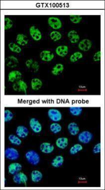
- Immunofluorescence analysis of paraformaldehyde-fixed A431, using HDAC1(GTX100513) antibody at 1:200 dilution.
- Submitted by
- GeneTex (provider)
- Main image
- Experimental details
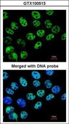
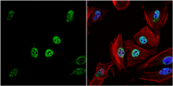
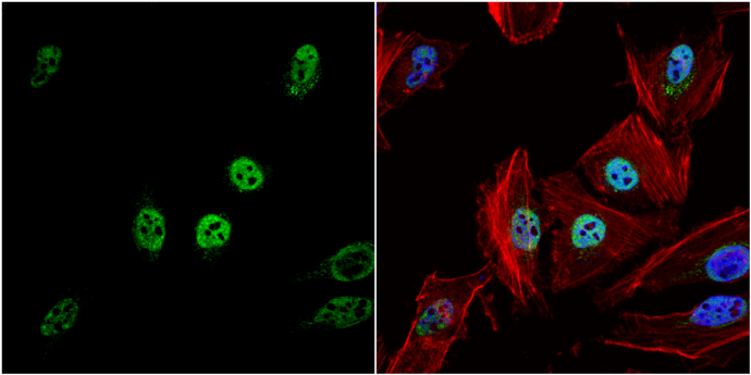
- HDAC1 antibody detects HDAC1 protein at nucleus by immunofluorescent analysis.Sample: HeLa cells were fixed in 4% paraformaldehyde at RT for 15 min.Green: HDAC1 protein stained by HDAC1 antibody (GTX100513) diluted at 1:500.Red: phalloidin, a cytoskeleton marker, stained by phalloidin (invitrogen, A12380) diluted at 1:200.Blue: Hoechst 33342 staining.
Supportive validation
- Submitted by
- GeneTex (provider)
- Main image
- Experimental details
- HDAC1antibody immunoprecipitates HDAC1 protein in IP experiments. IP Sample: 1000 £gg 293T whole cell lysate/extract A. 40 £gg 293T whole cell lysate/extract B. Control with 2.5 £gg of preimmune rabbit IgG C. Immunoprecipitation of HDAC1 protein by 2.5 £gg of HDAC1 antibody (GTX100513) 10% SDS-PAGE The immunoprecipitated HDAC1 protein was detected by HDAC1 antibody (GTX100513) diluted at 1:1000. EasyBlot anti-rabbit IgG (GTX221666-01) was used as a secondary reagent.
Supportive validation
- Submitted by
- GeneTex (provider)
- Main image

- Experimental details
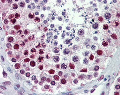
- Immunohistochemical analysis of paraffin-embedded human testis, using HDAC1(GTX100513) antibody(10 £gg/ml).
- Submitted by
- GeneTex (provider)
- Main image
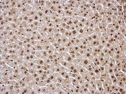
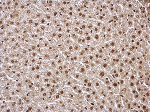
- Experimental details
- HDAC1 antibody detects HDAC1 protein at nucleus on mouse liver by immunohistochemical analysis. Sample: Paraffin-embedded mouse liver. HDAC1 antibody (GTX100513) dilution: 1:500.
- Submitted by
- GeneTex (provider)
- Main image

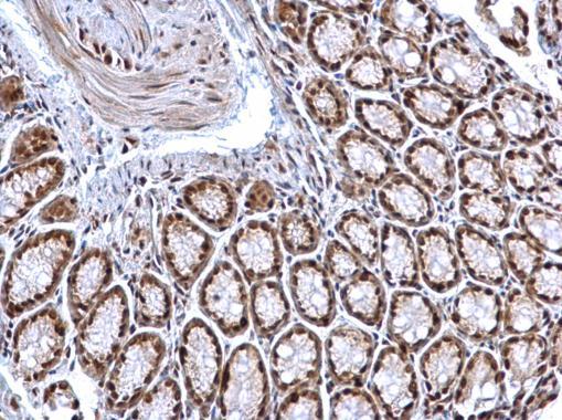
- Experimental details
- HDAC1 antibody detects HDAC1 protein at nucleus on mouse colon by immunohistochemical analysis. Sample: Paraffin-embedded mouse colon. HDAC1 antibody (GTX100513) dilution: 1:500.
- Submitted by
- GeneTex (provider)
- Main image
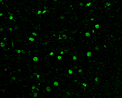
- Experimental details
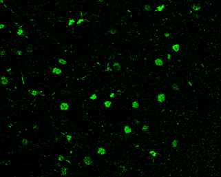
- HDAC1 antibody detects HDAC1 protein at nucleus in rat brain by immunohistochemical analysis. Sample: Paraffin-embedded rat brain. Green: HDAC1 antibody (GTX100513) diluted at 1:200. The signal was developed using goat anti-rabbit IgG antibody (Dylight488) (GTX213110-04).
- Submitted by
- GeneTex (provider)
- Main image
- Experimental details
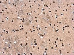
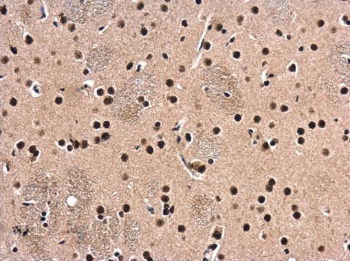
- HDAC1 antibody detects HDAC1 protein at cytoplasm and nucleus in rat brain by immunohistochemical analysis. Sample: Paraffin-embedded rat brain. HDAC1 antibody (GTX100513) diluted at 1:500.
- Submitted by
- GeneTex (provider)
- Main image

- Experimental details
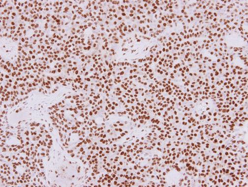
- HDAC1 antibody detects HDAC1 protein at nucleus in human lung adenocarcinoma by immunohistochemical analysis. Sample: Paraffin-embedded human lung adenocarcinoma. HDAC1 antibody (GTX100513) diluted at 1:250.
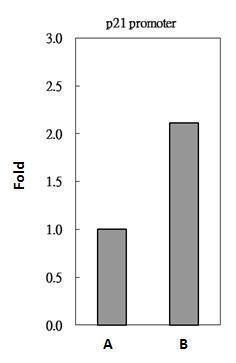
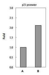
Supportive validation
- Submitted by
- GeneTex (provider)
- Main image
- Experimental details
- HDAC1 antibody immunoprecipitates HDAC1 protein-DNA in ChIP experiments. ChIP Sample: 293T whole cell lysate/extract A. 5 £gg preimmune rabbit IgG B. 5 £gg of HDAC1 antibody (GTX100513) The precipitated DNA was detected by PCR with primer set targeting to p21 promoter.