Explore
 Validate
 Learn
LearnMA5-18071
antibody from Invitrogen Antibodies
Targeting: HDAC1
GON-10, HD1, KDAC1, RPD3L1
 Western blot Immunocytochemistry
 Immunoprecipitation Immunohistochemistry Flow cytometry Chromatin Immunoprecipitation
Antibody data
- Antibody Data
- Antigen structure
- References [0]
- Comments [0]
- Validations
- Western blot [4]
- Immunocytochemistry [1]
- Chromatin Immunoprecipitation [1]
Submit
Validation data
Reference
Comment
Report error
- Product number
- MA5-18071 - Provider product page

- Provider
- Invitrogen Antibodies
- Product name
- HDAC1 Monoclonal Antibody (10E2)
- Antibody type
- Monoclonal
- Antigen
- Synthetic peptide
- Description
- This target has a predicted molecular weight of 55 kDa. Actual Western blot results show a band at 57 kDa.
- Reactivity
- Human, Mouse, Rat
- Host
- Mouse
- Isotype
- IgG
- Antibody clone number
- 10E2
- Vial size
- 100 µL
- Concentration
- 1 mg/mL
- Storage
- Maintain refrigerated at 2-8°C for up to 1 month. For long term storage store at -20°C
No comments: Submit comment
Supportive validation
- Submitted by
- Invitrogen Antibodies (provider)
- Main image
- Experimental details
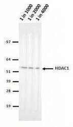
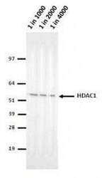
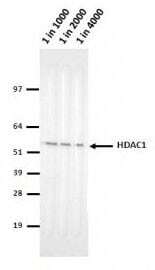
- Western blot analysis of HDAC1 in Hela lysates using an HDAC1 monoclonal antibody (Product # MA5-18071) at various dilutions, followed by detection using an AP-conjugated mouse IgG whole molecule antibody at a dilution of 1:2000 on a BCIP/NBT substrate.
- Submitted by
- Invitrogen Antibodies (provider)
- Main image
- Experimental details
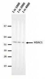
- Western blot analysis of HDAC1 in Hela lysates using an HDAC1 monoclonal antibody (Product # MA5-18071) at various dilutions, followed by detection using an AP-conjugated mouse IgG whole molecule antibody at a dilution of 1:2000 on a BCIP/NBT substrate.
- Submitted by
- Invitrogen Antibodies (provider)
- Main image

- Experimental details
- Knockdown of HDAC1 was achieved by transfecting MCF7 cells with HDAC1 specific siRNAs (Silencer® select Product # s73). Western blot analysis (Fig. a) was performed using Modified whole cell extracts (1% SDS) from the HDAC1 knockdown cells (Lane 3), non-specific scrambled siRNA transfected cells (Lane 2) and untransfected cells (Lane 1). The blot was probed with Anti-HDAC1 Monoclonal Antibody (10E2) (Product # MA5-18071, 1:1000 dilution) and Goat anti-Mouse IgG (H+L) Superclonal™ Recombinant Secondary Antibody, HRP (Product # A28177, 1:4000 dilution) using the iBright FL 1000 (Product # A32752). Densitometric analysis of this Western Blot is shown in histogram (Fig. b). Decrease in signal upon siRNA mediated knock down confirms that antibody is specific to HDAC1..

- Submitted by
- Invitrogen Antibodies (provider)
- Main image
- Experimental details
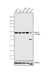
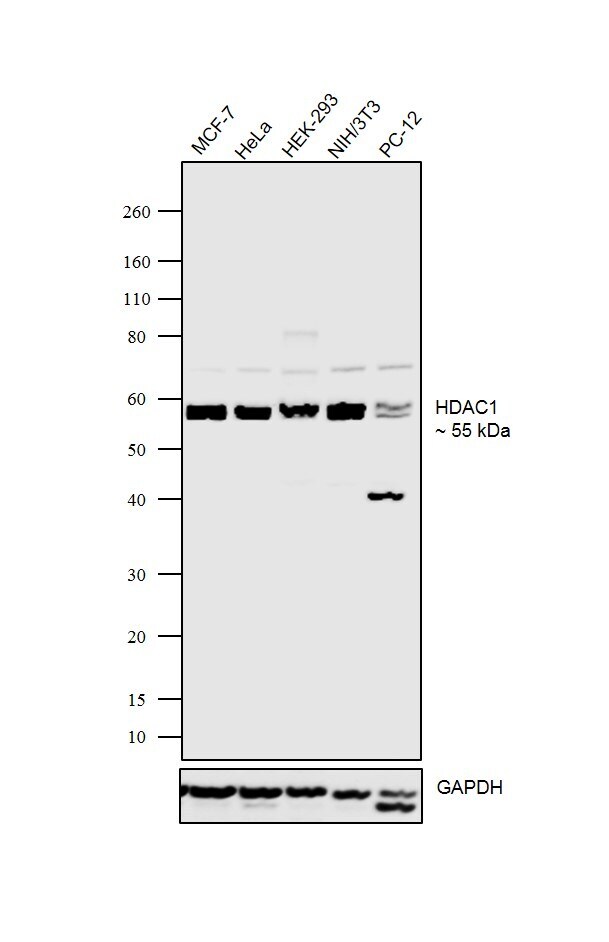
- Western blot was performed using Anti-HDAC1 Polyclonal Antibody (Product # MA5-18071) and ~55kDa band corresponding to HDAC1 was observed in MCF-7, HeLa, HEK-293, NIH/3T3 and PC-12 cells. Modified whole cell extracts (1% SDS) (30 ug lysate) of MCF-7 (Lane 1), HeLa (Lane 2), HEK-293 (Lane 3), NIH/3T3 (Lane 4) and PC-12 (Lane 5) were electrophoresed using NuPAGE® 4-12% Bis-Tris gel (Product # NP0322BOX). Resolved proteins were then transferred onto a nitrocellulose membrane (Product # IB23001) by iBlot® 2 Dry Blotting System (Product # IB21001).The blot was probed with the primary antibody (1:1000 dilution) and detected by chemiluminescence with Goat anti-Mouse IgG (H+L) Superclonal™ Recombinant Secondary Antibody, HRP (Product # A28177, 1:4000 dilution) using the iBright FL 1000 (Product # A32752). Chemiluminescent detection was performed using Novex® ECL Chemiluminescent Substrate Reagent Kit (Product # WP20005)..
Supportive validation
- Submitted by
- Invitrogen Antibodies (provider)
- Main image
- Experimental details
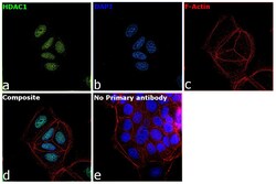
- Immunofluorescence analysis of HDAC1 was performed using 70% confluent log phase MCF7 cells. The cells were fixed with 4% paraformaldehyde for 10 minutes, permeabilized with 0.1% Triton™ X-100 for 15 minutes, and blocked with 2% BSA for 1 hour at room temperature. The cells were labeled with HDAC1 Monoclonal Antibody (10E2) (Product # MA5-18071) at 1 µg/mL in 0.1% BSA, incubated at 4 degree Celsius overnight and then labeled with Goat anti-Mouse IgG (H+L), Superclonal™ Recombinant Secondary Antibody, Alexa Fluor 488 (Product # A28175) at a dilution of 1:2000 for 45 minutes at room temperature (Panel a: green). Nuclei (Panel b: blue) were stained with SlowFade® Gold Antifade Mountant with DAPI (Product # S36938). F-actin (Panel c: red) was stained with Rhodamine Phalloidin (Product # R415, 1:300). Panel d represents the merged image showing nuclear localization. Panel e represents control cells with no primary antibody to assess background. The images were captured at 60X magnification.

Supportive validation
- Submitted by
- Invitrogen Antibodies (provider)
- Main image

- Experimental details
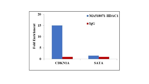
- Chromatin Immunoprecipitation (ChIP) assay of endogenous HDAC1 protein using Anti-HDAC1 Antibody: ChIP was performed using Anti-HDAC1 Mouse Monoclonal Antibody (Product # MA5-18071, 5 µg) on sheared chromatin from HeLa cells using the MAGnify ChIP System kit (Product # 49-2024). Normal Mouse IgG was used as a negative IP control. The purified DNA was analyzed by qPCR using primers binding to CDKN1A Intron 1 and SATA satellite repeats. Data is presented as fold enrichment of the antibody signal versus the negative control IgG using the comparative CT method.