Explore
 Validate
 Learn
Learn Western blot
 ELISA
ELISAAntibody data
- Antibody Data
- Antigen structure
- References [0]
- Comments [0]
- Validations
- ELISA [1]
- Immunocytochemistry [1]
- Immunoprecipitation [1]
- Chromatin Immunoprecipitation [1]
Submit
Validation data
Reference
Comment
Report error
- Product number
- GTX20012 - Provider product page

- Provider
- GeneTex
- Product name
- HDAC1 antibody
- Antibody type
- Polyclonal
- Reactivity
- Human, Mouse
- Host
- Rabbit
No comments: Submit comment
Supportive validation
- Submitted by
- GeneTex (provider)
- Main image

- Experimental details
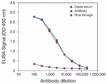
- To determine the titer, an ELISA was performed using anti-HDAC1 crude serum, purified anti-HDAC1 antibodies (GTX20012), and the column flow through obtained from the antibody purification step. The antigen used was the C-terminal peptide used for immunization. By plotting the absorbance against the antibody dilution, the titer of the antibody was estimated to be 1:4,250.
Supportive validation
- Submitted by
- GeneTex (provider)
- Main image
- Experimental details
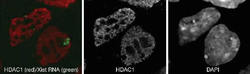
- Mouse differentiated ES cells were formaldehyde fixed, permeabilized with Triton? X-100 and then blocked with PBS containing BSA. Cells were labeled with the anti-HDAC1 (diluted 1:200 and incubated for 45 minutes at room temperature) followed by labeled goat anti-rabbit secondary antibody. Subsequently, RNA FISH (fluorescence in situ hybridization) was performed to detect Xist RNA (green signal). Nuclei were DAPI-stained to specifically label the DNA. As expected, both DAPI and HDAC1 staining occur in the nucleus of the cells. Note the broadly dispersed characteristic pattern of this protein mark in interphase chromatin but its exclusion from constitutive heterochromatin in mouse cells

Supportive validation
- Submitted by
- GeneTex (provider)
- Main image
- Experimental details
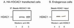
- A. Immunoprecipitation of HDAC1 from transfected cells. HEK293T cells were transiently transfected with an expression vector for HA-tagged HDAC1. Immunoprecipitations were performed on cell extracts with 2 ?g of the anti-HDAC1 antibody (GTX20012) (lane 2). As an IP positive control, an anti-HA antibody (lane 3), and, as negative control, beads only (lane 1) were used. The presence of HA-HDAC1 in the immunoprecipitates was detected by western blot analysis using the anti-HA antibody. B. Immunoprecipitation of endogenous HDAC1 from untransfected cells. Immunoprecipitations were performed on HeLa extracts with the anti-HDAC1 antibody (GTX20012). The presence of HDAC1 in the immunoprecipitates (lane 5) and in the HeLa extracts (lane 6) was detected by western blot analysis with the HDAC1 antibody. Beads only were used as negative control (lane 4).

Supportive validation
- Submitted by
- GeneTex (provider)
- Main image
- Experimental details
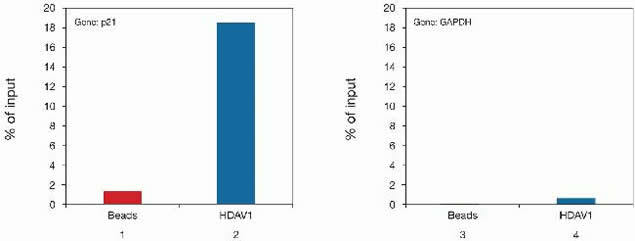
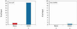
- ChIP assays were performed using U-2OS cells, the anti-HDAC1 antibody (GTX20012), and optimized PCR primer sets for qPCR. Each ChIP assay used sheared chromatin from 1 million cells and 2.4 ?g of anti-HDAC1 antibody. The results, expressed as the relative amount of immunoprecipitated DNA compared to input DNA (% of input) are shown. Left: ChIP results using the anti-HDAC1 antibody (bar 2) or beads only (bar 1) and PCR primers specific for p21. The occupancy of p21 (a known HDAC1 target gene) by HDAC1 is clearly demonstrated. Right: ChIP results using the anti-HDAC1 antibody (bar 4) or beads only (bar 3) and PCR primers for GAPDH (used as negative control).