Explore
 Validate
 Learn
LearnNBP1-31294
antibody from Novus Biologicals
Targeting: HDAC1
GON-10, HD1, KDAC1, RPD3L1
 Western blot
 Immunocytochemistry Immunoprecipitation Immunohistochemistry Chromatin Immunoprecipitation
Antibody data
- Antibody Data
- Antigen structure
- References [0]
- Comments [0]
- Validations
- Western blot [7]
- Immunohistochemistry [6]
- Chromatin Immunoprecipitation [1]
Submit
Validation data
Reference
Comment
Report error
- Product number
- NBP1-31294 - Provider product page

- Provider
- Novus Biologicals
- Proper citation
- Novus Cat#NBP1-31294, RRID:AB_10003773
- Product name
- Rabbit Polyclonal HDAC1 Antibody
- Antibody type
- Polyclonal
- Description
- Immunogen affinity purified.
- Reactivity
- Human, Mouse, Rat
- Host
- Rabbit
- Isotype
- IgG
- Vial size
- 100 ul
- Storage
- Aliquot and store at -20C or -80C. Avoid freeze-thaw cycles.
No comments: Submit comment
Supportive validation
- Submitted by
- Novus Biologicals (provider)
- Main image
- Experimental details
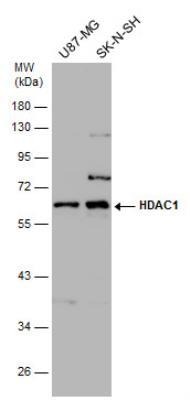
- Western Blot: HDAC1 Antibody [NBP1-31294] - NIH-3T3 7.5% SDS PAGE diluted at 1:1000
- Submitted by
- Novus Biologicals (provider)
- Main image

- Experimental details
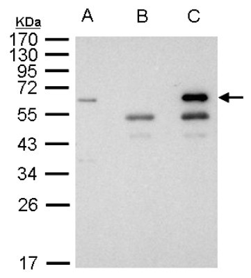
- Western Blot: HDAC1 Antibody [NBP1-31294] - Sample: 1000 ug 293T whole cell lysate/extract A. 40 ug 293T whole cell lysate/extract, B. Control with 2. 5 ug of preimmune rabbit IgG, C. Immunoprecipitation of HDAC1 protein by 2. 5 ug of HDAC1 antibody 10% SDS-PAGE gel.
- Submitted by
- Novus Biologicals (provider)
- Main image
- Experimental details
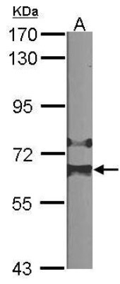
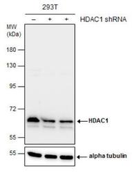
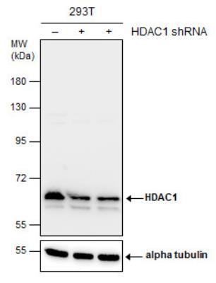
- Western Blot: HDAC1 Antibody [NBP1-31294] - Non-transfected (-) and transfected (+) 293T whole cell extracts (30 ug) were separated by 7.5% SDS-PAGE, and the membrane was blotted with HDAC1 antibody. The HRP-conjugated anti-rabbit IgG antibody was used to detect the primary antibody.
- Submitted by
- Novus Biologicals (provider)
- Main image

- Experimental details
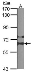
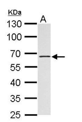
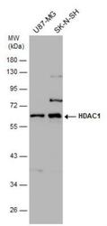
- Western Blot: HDAC1 Antibody [NBP1-31294] - A. 30 ug Rat2 whole cell lysate/extract 10% SDS-PAGE HDAC1 antibody dilution: 1:1000 The HRP-conjugated anti-rabbit IgG antibody (NBP2-19301) was used to detect the primary antibody.
- Submitted by
- Novus Biologicals (provider)
- Main image
- Experimental details
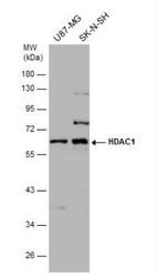
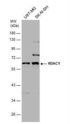
- Western Blot: HDAC1 Antibody [NBP1-31294] - Various whole cell extracts (30 ug) were separated by 10% SDS-PAGE, and the membrane was blotted with HDAC1 antibody diluted at 1:1000. The HRP-conjugated anti-rabbit IgG antibody (NBP2-19301) was used to detect the primary antibody.
- Submitted by
- Novus Biologicals (provider)
- Main image
- Experimental details
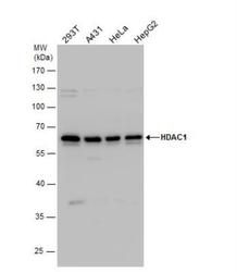
- Western Blot: HDAC1 Antibody [NBP1-31294] - Various whole cell extracts (30 ug) were separated by 10% SDS-PAGE, and the membrane was blotted with HDAC1 antibody diluted by 1:1000. The HRP-conjugated anti-rabbit IgG antibody (NBP2-19301) was used to detect the primary antibody.
- Submitted by
- Novus Biologicals (provider)
- Main image
- Experimental details
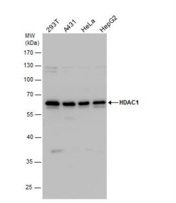
- Western Blot: HDAC1 Antibody [NBP1-31294] - Various whole cell extracts (30 ug) were separated by 10% SDS-PAGE, and the membrane was blotted with HDAC1 antibody diluted at 1:1000. The HRP-conjugated anti-rabbit IgG antibody was used to detect the primary antibody.
Supportive validation
- Submitted by
- Novus Biologicals (provider)
- Main image
- Experimental details
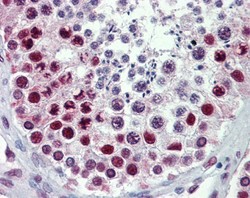
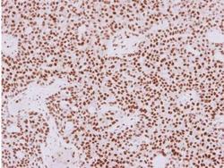
- Immunohistochemistry-Paraffin: HDAC1 Antibody [NBP1-31294] - Human testis, using HDAC1 antibody(10 ug/ml). Antigen Retrieval: Trilogy™ (EDTA based, pH 8.0) buffer, 15min.

- Submitted by
- Novus Biologicals (provider)
- Main image

- Experimental details
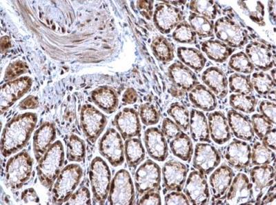
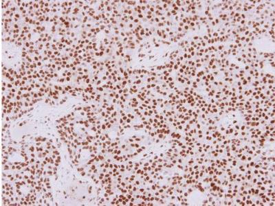
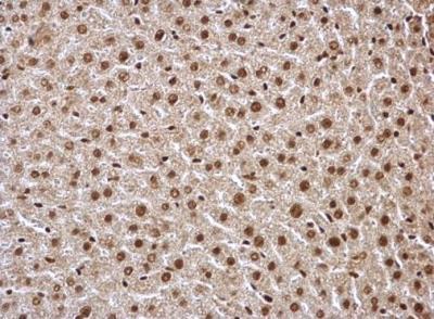
- Immunohistochemistry-Paraffin: HDAC1 Antibody [NBP1-31294] - Mouse colon. HDAC1 antibody dilution: 1:500. Antigen Retrieval: Trilogy™ (EDTA based, pH 8.0) buffer, 15min.
- Submitted by
- Novus Biologicals (provider)
- Main image
- Experimental details
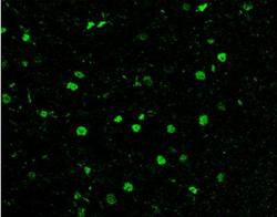
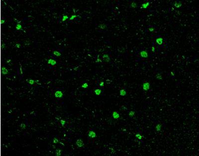
- Immunohistochemistry-Paraffin: HDAC1 Antibody [NBP1-31294] - Rat brain. Green: HDAC1 antibody diluted at 1:200. The signal was developed using goat anti-rabbit IgG antibody (Dylight488). Antigen Retrieval: Citrate buffer, pH 6.0, 15 min.
- Submitted by
- Novus Biologicals (provider)
- Main image
- Experimental details
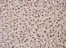
- Immunohistochemistry-Paraffin: HDAC1 Antibody [NBP1-31294] - Sample: Paraffin-embedded mouse liver. HDAC1 antibody dilution: 1:500. Antigen Retrieval: Trilogy™ (EDTA based, pH 8.0) buffer, 15min.
- Submitted by
- Novus Biologicals (provider)
- Main image
- Experimental details
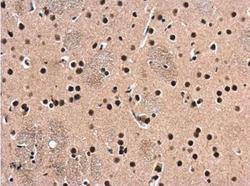
- Immunohistochemistry-Paraffin: HDAC1 Antibody [NBP1-31294] - Rat brain. HDAC1 antibody diluted at 1:500. Antigen Retrieval: Citrate buffer, pH 6.0, 15 min.

- Submitted by
- Novus Biologicals (provider)
- Main image
- Experimental details
- Immunohistochemistry-Paraffin: HDAC1 Antibody [NBP1-31294] - Mouse liver. HDAC1 antibody dilution: 1:500. Antigen Retrieval: Trilogy™ (EDTA based, pH 8.0) buffer, 15min.
Supportive validation
- Submitted by
- Novus Biologicals (provider)
- Main image
- Experimental details
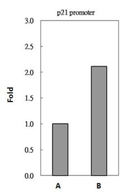
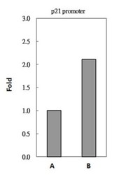
- Chromatin Immunoprecipitation: HDAC1 Antibody [NBP1-31294] - Sample: 293T whole cell lysate/extract A. 5 ug preimmune rabbit IgG, B. 5 ug of HDAC1 antibody The precipitated DNA was detected by PCR with primer set targeting to p21 promoter.