 Explore
 Validate
 Learn
Learn Western blot
 ELISA
ELISAAntibody data
- Antibody Data
- Antigen structure
- References [0]
- Comments [0]
- Validations
- Western blot [1]
- Immunohistochemistry [2]
Submit
Validation data
Reference
Comment
Report error
- Product number
- AP09288PU-N - Provider product page

- Provider
- Acris Antibodies GmbH
- Proper citation
- Acris Antibodies GmbH Cat#AP09288PU-N, RRID:AB_2035620
- Product name
- anti HDAC1 (466-482)
- Antibody type
- Polyclonal
- Antigen
- Synthetic peptide corresponding to amino acids 466-482 of Human HDAC-1
- Reactivity
- Human, Mouse, Rat, Bovine, Chicken/Avian
- Host
- Rabbit
- Isotype
- IgG
- Vial size
- 0.1 mg
- Concentration
- 1.33 mg/ml (by UV absorbance at 280 nm)
No comments: Submit comment
Supportive validation
- Submitted by
- Acris Antibodies GmbH (provider)
- Main image

- Experimental details
- Western blot using Affinity Purified anti-HDAC-1 antibody shows detection of a band at ~65 kDa corresponding to human HDAC1 present in a 293 whole cell lysate (arrowhead). Approximately 35 µg of lysate wseparated on a 4-20% Tris-HEPES gel by SDS-PAGE and transferred onto nitrocellulose. After blocking the membrane was probed with the primary antibody diluted to 1:1,350. Reaction occurred 2 h at room temperature followed by washes and reaction with a 1:10,000 dilution of IRDyeas (TM)800 conjugated Rb-a-Goat IgG [H&L] MXHu for 45 min at room temperature. IRDye(TM)800 fluorescence image was captured using the Odyssey(R) Infrared Imaging System developed by LI-COR. IRDye is a trademark of LI-COR, Inc. Other detection systems will yield similar results.

Supportive validation
- Submitted by
- Acris Antibodies GmbH (provider)
- Main image
- Experimental details
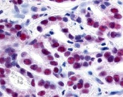
- Immunohistochemistry. Affinity Purified anti-HDAC-1 antibody was used at a 1:500 dilution to detect HDAC-1 by immunohistochemistry in human prostate cancer tissue. Tissue was formalin-fixed and paraffin embedded.

- Submitted by
- Acris Antibodies GmbH (provider)
- Main image

- Experimental details
- Immunohistochemistry. Affinity Purified anti-HDAC1 antibody shows strong nuclear staining of tumor cells in human lung tissue. Tissue was formalin-fixed and paraffin embedded. Brown color indicates presence of protein, blue color shows cell nuclei.
