 Explore
 Validate
 Learn
Learn Western blot
 Immunoprecipitation
Antibody data
- Antibody Data
- Antigen structure
- References [1]
- Comments [0]
- Validations
- Western blot [4]
- Immunocytochemistry [1]
- Immunohistochemistry [1]
- Other assay [1]
Submit
Validation data
Reference
Comment
Report error
- Product number
- MA1-23202 - Provider product page

- Provider
- Invitrogen Antibodies
- Product name
- E2F1 Monoclonal Antibody (17E2)
- Antibody type
- Monoclonal
- Antigen
- Other
- Description
- A suggested positive control is E2F-1 transfected Soas2 cells.
- Reactivity
- Human, Mouse, Rat
- Host
- Mouse
- Isotype
- IgG
- Antibody clone number
- 17E2
- Vial size
- 100 µL
- Concentration
- 1 mg/mL
- Storage
- Store at 4°C short term. For long term storage, store at -20°C, avoiding freeze/thaw cycles.
Submitted references PRR11 promotes cell proliferation by regulating PTTG1 through interacting with E2F1 transcription factor in pan-cancer.
Zhang H, He Z, Qiu L, Wei J, Gong X, Xian M, Chen Z, Cui Y, Fu S, Zhang Z, Hu B, Zhang X, Lin S, Du H
Frontiers in molecular biosciences 2022;9:877320
Frontiers in molecular biosciences 2022;9:877320
No comments: Submit comment
Supportive validation
- Submitted by
- Invitrogen Antibodies (provider)
- Main image

- Experimental details
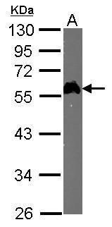
- Western blot analysis of Transcription Factor E2F1 using 50 µg of mouse spleen lysate. Samples were loaded onto a 10% SDS-PAGE gel and probed with a Transcription Factor E2F1 monoclonal antibody (Product # MA1-23202) at a dilution of 1:1000.
- Submitted by
- Invitrogen Antibodies (provider)
- Main image
- Experimental details
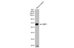
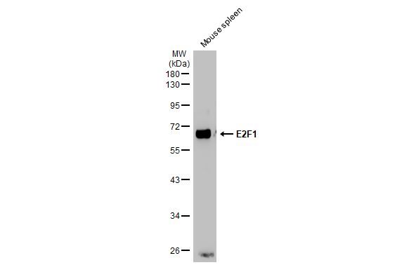
- Western Blot using E2F1 Monoclonal Antibody (17E2) (Product # MA1-23202). Mouse tissue extract (50 µg) was separated by 10% SDS-PAGE, and the membrane was blotted with E2F1 Monoclonal Antibody (17E2) (Product # MA1-23202) diluted at 1:1,000. The HRP-conjugated anti-mouse IgG antibody was used to detect the primary antibody.
- Submitted by
- Invitrogen Antibodies (provider)
- Main image
- Experimental details
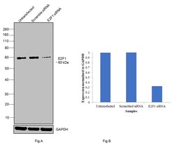
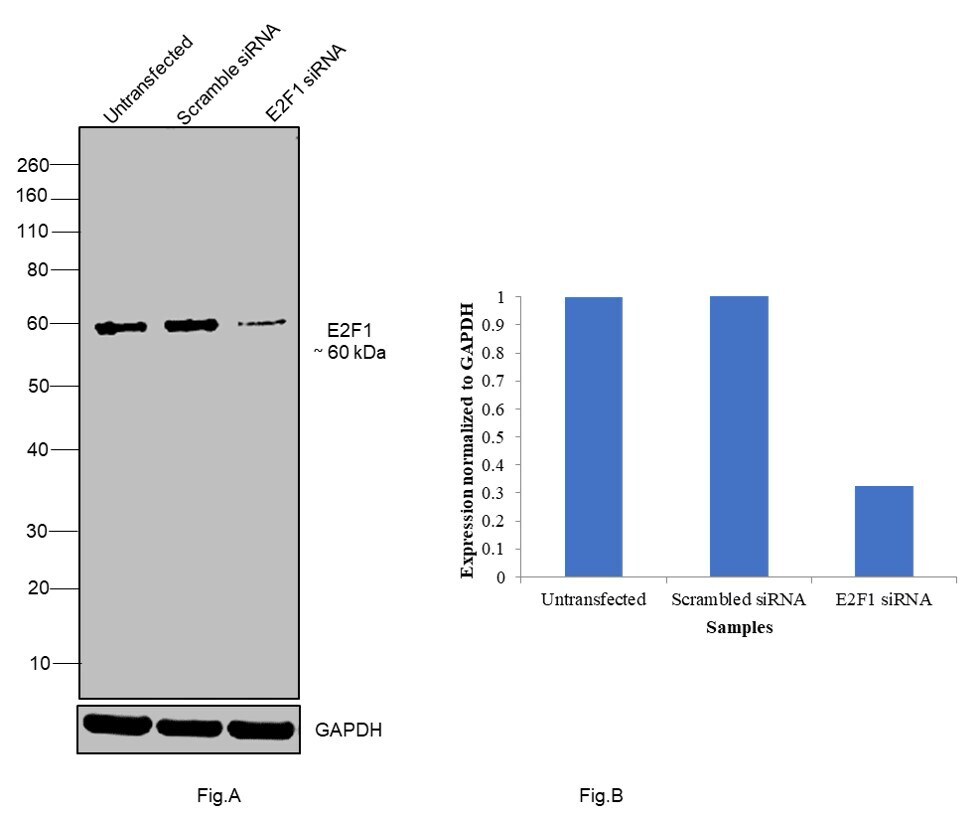
- Knockdown of E2F1 was achieved by transfecting Hep G2 with E2F1 specific siRNA (Silencer® select Product # s4405). Western blot analysis (Fig. a) was performed using nuclear enriched extracts from the untransfected cells (Lane 1), non-specific scrambled siRNA transfected cells (Lane 2) and E2F1 knockdown cells (Lane 3). The blot was probed with E2F1 Monoclonal Antibody (17E2) (Product # MA1-23202, 1:1000 dilution) and Goat anti-Mouse IgG (H+L) Superclonal™ Recombinant Secondary Antibody, HRP conjugate (Product # A28177, 0.25 µg/mL, 1:4000 dilution). Densitometric analysis of this western blot is shown i histogram (Fig. b). Loss of signal upon siRNA mediated knock down confirms that antibody is specific to E2F1.
- Submitted by
- Invitrogen Antibodies (provider)
- Main image
- Experimental details
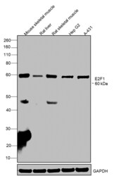
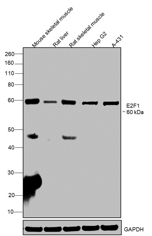
- Western blot was performed using E2F1 Monoclonal Antibody (17E2) (Product # MA1-23202) and a 60 kDa band corresponding to E2F1 was observed across cell lines tested. Whole cell extracts (30 µg lysate) of Mouse skeletal muscle (Lane 1), Rat liver (Lane 2), Rat skeletal muscle (Lane 3), Hep G2 (Lane 4) and A-431 (Lane 5) were electrophoresed using NuPAGE® 4-12 % Bis-Tris gel (Product # NP0321BOX). Resolved proteins were then transferred onto a nitrocellulose membrane (Product # IB23001) by iBlot® 2 Dry Blotting System (Product # IB21001). The blot was probed with the primary antibody (1:1000 dilution) and detected by chemiluminescence with Goat anti-Mouse IgG (H+L), Superclonal™ Recombinant Secondary Antibody, HRP (Product # A28177, 1:4000 dilution) using the iBright FL 1000 (Product # A32752). Chemiluminescent detection was performed using Novex® ECL Chemiluminescent Substrate Reagent Kit (Product # WP20005).
Supportive validation
- Submitted by
- Invitrogen Antibodies (provider)
- Main image
- Experimental details
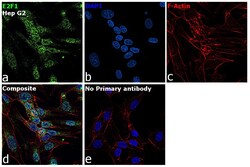
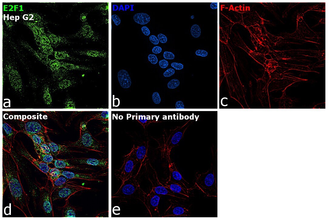
- Immunofluorescence analysis of E2F1 was performed using 70% confluent log phase Hep G2 cells. The cells were fixed with 4% paraformaldehyde for 10 minutes, permeabilized with 0.1% Triton™ X-100 for 15 minutes, and blocked with 2% BSA for 1 hour at room temperature. The cells were labeled with E2F1 Monoclonal Antibody (17E2) (Product # MA1-23202) at 5 µg/mL in 0.1% BSA, incubated at 4 degree Celsius overnight and then labeled with Goat anti-Rabbit IgG (H+L) Superclonal™ Recombinant Secondary Antibody, Alexa Fluor® 488 conjugate (Product # A27034) at a dilution of 1:2000 for 45 minutes at room temperature (Panel a: green). Nuclei (Panel b: blue) were stained with SlowFade® Gold Antifade Mountant with DAPI (Product # S36938). F-actin (Panel c: red) was stained with Rhodamine Phalloidin (Product # R415, 1:300). Panel d represents the merged image showing localization to nucleus. Panel f represents control cells with no primary antibody to assess background. The images were captured at 60X magnification.
Supportive validation
- Submitted by
- Invitrogen Antibodies (provider)
- Main image
- Experimental details
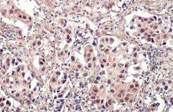
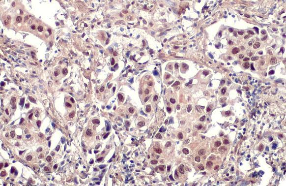
- E2F1 Monoclonal Antibody (17E2) detects E2F1 protein at nucleus by immunohistochemical analysis. Sample: Paraffin-embedded human lung cancer. E2F1 stained by E2F1 Monoclonal Antibody (17E2) (Product # MA1-23202) diluted at 1:100. Antigen Retrieval: Citrate buffer, pH 6.0, 15 min.
Supportive validation
- Submitted by
- Invitrogen Antibodies (provider)
- Main image
- Experimental details
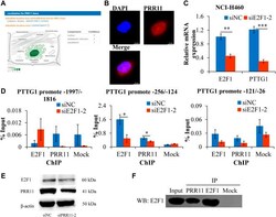
- FIGURE 6 PRR11 promotes the expression level of PTTG1 by interacting with E2F1. (A) Localization for PRR11 gene was analyzed using GeneCards ( https://www.genecards.org/ ). (B) Immunofluorescence localization of PRR11. (C) Expression levels of PTTG1 after interfering E2F1. (D) ChIP-qPCR of the PTTG1 promoter region. (E) Expression levels of E2F1 and PRR11 in NCI-H460 cells transfected with siNC or siPRR11 were examined by Western blotting. (F) Co-IP of PRR11 and E2F1 in NCI-H460 cells. N = 3. * p < 0.05, ** p < 0.01, and *** p < 0.001.
