Explore
 Validate
 Learn
Learn Western blot
Antibody data
- Antibody Data
- Antigen structure
- References [0]
- Comments [0]
- Validations
- Western blot [1]
- Immunohistochemistry [1]
Submit
Validation data
Reference
Comment
Report error
- Product number
- TA319248 - Provider product page

- Provider
- OriGene
- Product name
- Rabbit polyclonal E2F-1 phospho S364 antibody
- Antibody type
- Polyclonal
- Description
- Rabbit polyclonal E2F-1 phospho S364 antibody
- Host
- Rabbit
- Conjugate
- Unconjugated
- Epitope
- E2F1
- Isotype
- IgG
- Antibody clone number
- NULL
- Vial size
- 100 µg
- Concentration
- 1.0 mg/mL
No comments: Submit comment
Supportive validation
- Submitted by
- OriGene (provider)
- Main image

- Experimental details
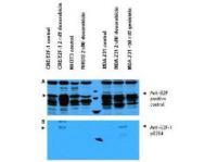
- WB using E2F-1 pS364 ab shows detection of a ~47 kDa band corresponding to phosphorylated E2F-1 in induced cell lysates. Panel A shows reactivity using a control ab reactive to all forms of E2F (arrowheads). Panel B shows specific reactivity against phosphorylated E2F-1 (arrowheads) using anti-E2F-1 pS364. Lysates are as follows: CRE/E2F-1 are CRE cells derived from mouse NIH3T3 line transfected with human E2F-1, NIH-3T3 used as a negative control, and MDA-MB-231 cells are a human breast cancer line.?Lysate was prepared from untreated cells or cells treated with 2 uM Doxorubicin used as DNA damaging agent. MDA-MB-231 cells were also treated with genistein, a mild DNA damaging agent.?The figure shows the same membrane first probed with the anti-E2F-1 pS364 at 1:250, then stripped and re-probed with the pan E2F ab used as a positive control. The positive control ab shows an E2F-1 band in all human cell lines, but not mouse cells. Treatment with doxorubicin increases the expression of E2F-1 as shown in Panel A. After film development, images were overlapped to confirm that specific anti-E2F-1 pS364 staining shown treated human cells in Panel B specifically aligns with E2F-1 staining shown in Panel A.
- Validation comment
- WB
Supportive validation
- Submitted by
- OriGene (provider)
- Main image
- Experimental details
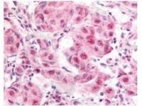
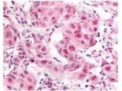
- Anti- E2F-1 pS364 antibody was used at a 10 ug/ml to detect nuclear and occasion-ally cytoplasmic signal in a variety of tissues including multi-human, multi-brain and multi-cancer slides. Within the multi-tumor block, the antibody showed variable levels of nuclear staining between individual tumors, with some tumors showing strong staining. This image shows E2F-1 pS364 staining of human breast carcinoma. Tissue was formalin-fixed and paraffin embedded.
- Validation comment
- IHC