Explore
 Validate
 Learn
Learn Western blot
Antibody data
- Antibody Data
- Antigen structure
- References [3]
- Comments [0]
- Validations
- Western blot [1]
- Immunohistochemistry [1]
Submit
Validation data
Reference
Comment
Report error
- Product number
- PAB10004 - Provider product page

- Provider
- Abnova Corporation
- Proper citation
- Abnova Corporation Cat#PAB10004, RRID:AB_1707637
- Product name
- E2F1 (phospho S364) polyclonal antibody
- Antibody type
- Polyclonal
- Description
- Rabbit polyclonal antibody raised against synthetic phosphopeptide of E2F1.
- Storage
- Store at 4°C. For long term storage store at -20°C.Aliquot to avoid repeated freezing and thawing.
Submitted references Induction of human metallothionein 1G promoter by VEGF and heavy metals: differential involvement of E2F and metal transcription factors.
Activation of p27Kip1 Expression by E2F1. A negative feedback mechanism.
The central acidic domain of MDM2 is critical in inhibition of retinoblastoma-mediated suppression of E2F and cell growth.
Joshi B, Ordonez-Ercan D, Dasgupta P, Chellappan S
Oncogene 2005 Mar 24;24(13):2204-17
Oncogene 2005 Mar 24;24(13):2204-17
Activation of p27Kip1 Expression by E2F1. A negative feedback mechanism.
Wang C, Hou X, Mohapatra S, Ma Y, Cress WD, Pledger WJ, Chen J
The Journal of biological chemistry 2005 Apr 1;280(13):12339-43
The Journal of biological chemistry 2005 Apr 1;280(13):12339-43
The central acidic domain of MDM2 is critical in inhibition of retinoblastoma-mediated suppression of E2F and cell growth.
Sdek P, Ying H, Zheng H, Margulis A, Tang X, Tian K, Xiao ZX
The Journal of biological chemistry 2004 Dec 17;279(51):53317-22
The Journal of biological chemistry 2004 Dec 17;279(51):53317-22
No comments: Submit comment
Supportive validation
- Submitted by
- Abnova Corporation (provider)
- Main image
- Experimental details
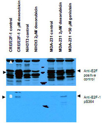
- Western blot using E2F1 (phospho S364) polyclonal antibody (Cat # PAB10004) shows detection of a band at ~47 kDa corresponding to phospho-E2F1 in induced cell lysates.Panel A shows reactivity using a control antibody reactive to all forms of E2F (arrowheads).Panel B shows specific reactivity against phosphorylated E2F-1 (arrowheads) using our Phospho-E2F1 S364 polyclonal antibody.Lysates are as follows : CRE/E2F1 are CRE cells derived from mouse NIH/3T3 line transfected with human E2F1, NIH/3T3 used as a negative control, and MDA-MB-231 cells are a human breast cancer line.As indicated each lysate was prepared from untreated cells and cells treated with 2 uM Doxorubicin used as a DNA damaging agent.In addition the MDA-MB-231 cells were also treated with genistein, a mild Ddamaging agent.The figure shows the same membrane first probed with the E2F1 (phospho S364) polyclonal antibody (Cat # PAB10004) used at a 1 : 250 dilution, then stripped and re-probed with the pan E2F antibody used as a positive control.The positive control antibody clearly shows an E2F1 band in all human cell lines, but not mouse cells.Treatment with doxorubicin increases the expression E2F1 as shown in Panel A.After film development, images were overlapped to confirm that specific anti-E2F1 pS364 staining shown treated human cells in Panel B specifically aligns with E2F1 staining shown in Panel A.Blots can be processed with HRP conjugated Gt-a-Rabbit IgG MX10 for 45 min at room temperature for ECL detection.Personal Communication, XiaoHe Yang, Univ. Oklahoma.

Supportive validation
- Submitted by
- Abnova Corporation (provider)
- Main image
- Experimental details
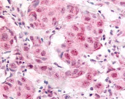
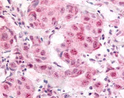
- Immunohistochemistry of E2F1 (phospho S364) polyclonal antibody (Cat # PAB10004) was used at a 10 ug/mL to detect nuclear and occasion-ally cytoplasmic signal in a variety of tissues in-cluding multi-human, multi-brain and multi-cancer slides.Within the multi-tumor block, the antibody showed variable levels of nuclear staining between individual tumors, with some tumors showing strong staining.This image shows E2F1 pS364 staining of human breast carcinoma. Tissue was formalin-fixed and paraffin embedded.Personal Communication, Tina Roush, Life Span Biosciences, Seattle, WA.
- Validation comment
- Immunohistochemistry (Formalin/PFA-fixed paraffin-embedded sections)