Explore
 Validate
 Learn
Learn Western blot
Antibody data
- Antibody Data
- Antigen structure
- References [1]
- Comments [0]
- Validations
- Western blot [1]
- Immunohistochemistry [1]
Submit
Validation data
Reference
Comment
Report error
- Product number
- AF4825 - Provider product page

- Provider
- R&D Systems
- Product name
- Human E2F-1 Antibody
- Antibody type
- Polyclonal
- Description
- Immunogen affinity purified. Detects human E2F-1 in direct ELISAs and Western blots. In direct ELISAs, less than 1% cross-reactivity with recombinant human E2F-2, -3, -4, -5, -6, -7, and -8 is observed.
- Reactivity
- Human
- Host
- Sheep
- Conjugate
- Unconjugated
- Antigen sequence
Q01094- Isotype
- IgG
- Vial size
- 100 ug
- Concentration
- LYOPH
- Storage
- Use a manual defrost freezer and avoid repeated freeze-thaw cycles. 12 months from date of receipt, -20 to -70 °C as supplied. 1 month, 2 to 8 °C under sterile conditions after reconstitution. 6 months, -20 to -70 °C under sterile conditions after reconstitution.
Submitted references A Cre-conditional MYCN-driven neuroblastoma mouse model as an improved tool for preclinical studies.
Althoff K, Beckers A, Bell E, Nortmeyer M, Thor T, Sprüssel A, Lindner S, De Preter K, Florin A, Heukamp LC, Klein-Hitpass L, Astrahantseff K, Kumps C, Speleman F, Eggert A, Westermann F, Schramm A, Schulte JH
Oncogene 2015 Jun;34(26):3357-68
Oncogene 2015 Jun;34(26):3357-68
No comments: Submit comment
Supportive validation
- Submitted by
- R&D Systems (provider)
- Main image

- Experimental details
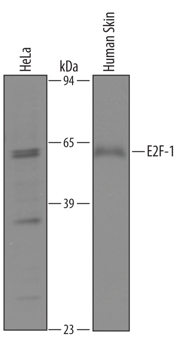
- Detection of Human E2F-1 by Western Blot. Western blot shows lysates of HeLa human cervical epithelial carcinoma cell line and human skin tissue. PVDF membrane was probed with 1 µg/mL of Sheep Anti-Human E2F-1 Antigen Affinity-purified Polyclonal Antibody (Catalog # AF4825) followed by HRP-conjugated Anti-Sheep IgG Secondary Antibody (Catalog # HAF016). A specific band was detected for E2F-1 at approximately 60 kDa (as indicated). This experiment was conducted under reducing conditions and using Immunoblot Buffer Group 8.
Supportive validation
- Submitted by
- R&D Systems (provider)
- Main image
- Experimental details
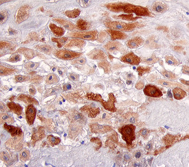
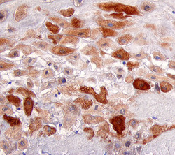
- E2F-1 in Human Placenta. E2F-1 was detected in immersion fixed paraffin-embedded sections of human placenta using 5 µg/mL Sheep Anti-Human E2F-1 Antigen Affinity-purified Polyclonal Antibody (Catalog # AF4825) overnight at 4 °C. Tissue was stained with the Anti-Sheep HRP-DAB Cell & Tissue Staining Kit (brown; Catalog # CTS019) and counterstained with hematoxylin (blue). View our protocol for Chromogenic IHC Staining of Paraffin-embedded Tissue Sections.