Explore
 Validate
 Learn
Learn Western blot
 Immunocytochemistry
Antibody data
- Antibody Data
- Antigen structure
- References [4]
- Comments [0]
- Validations
- Western blot [1]
Submit
Validation data
Reference
Comment
Report error
- Product number
- AF819 - Provider product page

- Provider
- Novus Biologicals
- Product name
- Rabbit Polyclonal Bad Antibody
- Antibody type
- Polyclonal
- Description
- Antigen Affinity-purified. Detects human and mouse Bad in Western blots.
- Reactivity
- Human, Mouse
- Host
- Rabbit
- Conjugate
- Unconjugated
- Isotype
- IgG
- Vial size
- 100 ug
- Concentration
- LYOPH
- Storage
- Use a manual defrost freezer and avoid repeated freeze-thaw cycles. 12 months from date of receipt, -20 to -70 degreesC as supplied. 1 month, 2 to 8 degreesC under sterile conditions after reconstitution. 6 months, -20 to -70 degreesC under sterile conditions after reconstitution.
Submitted references TCDD promotes lung tumors via attenuation of apoptosis through activation of the Akt and ERK1/2 signaling pathways.
Translocation of full-length Bid to mitochondria during anoikis.
Sustained early growth response gene 3 expression inhibits the survival of CD4/CD8 double-positive thymocytes.
Sustained early growth response gene 3 expression inhibits the survival of CD4/CD8 double-positive thymocytes.
Chen RJ, Siao SH, Hsu CH, Chang CY, Chang LW, Wu CH, Lin P, Wang YJ
PloS one 2014;9(6):e99586
PloS one 2014;9(6):e99586
Translocation of full-length Bid to mitochondria during anoikis.
Valentijn AJ, Gilmore AP
The Journal of biological chemistry 2004 Jul 30;279(31):32848-57
The Journal of biological chemistry 2004 Jul 30;279(31):32848-57
Sustained early growth response gene 3 expression inhibits the survival of CD4/CD8 double-positive thymocytes.
Xi H, Kersh GJ
Journal of immunology (Baltimore, Md. : 1950) 2004 Jul 1;173(1):340-8
Journal of immunology (Baltimore, Md. : 1950) 2004 Jul 1;173(1):340-8
Sustained early growth response gene 3 expression inhibits the survival of CD4/CD8 double-positive thymocytes.
Xi H, Kersh GJ
Journal of immunology (Baltimore, Md. : 1950) 2004 Jul 1;173(1):340-8
Journal of immunology (Baltimore, Md. : 1950) 2004 Jul 1;173(1):340-8
No comments: Submit comment
Supportive validation
- Submitted by
- Novus Biologicals (provider)
- Main image

- Experimental details
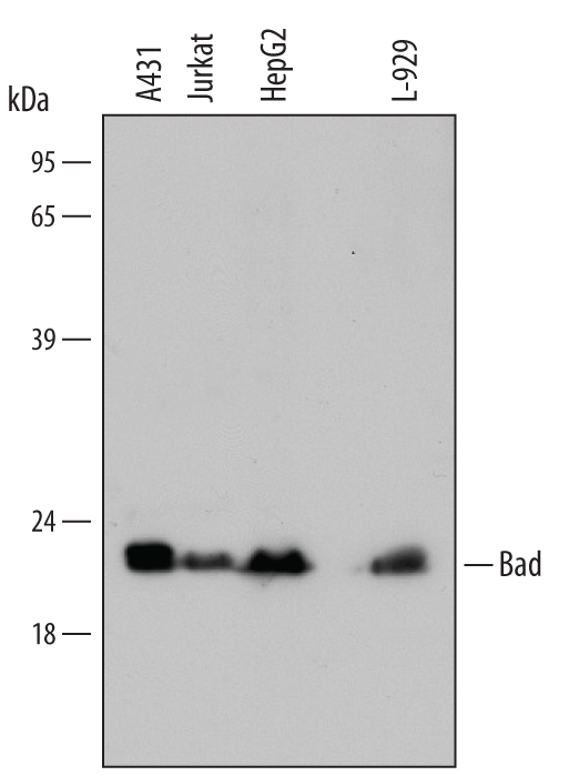
- Detection of Human/Mouse Bad by Western Blot. Western blot shows lysates of A431 human epithelial carcinoma cell line, Jurkat human acute T cell leukemia cell line, HepG2 human hepatocellular carcinoma cell line, and L-929 mouse fibroblast cell line. PVDF membrane was probed with 1 µg/mL of Rabbit Anti-Human/Mouse Bad Antigen Affinity-purified Polyclonal Antibody (Catalog # AF819) followed by HRP-conjugated Anti-Rabbit IgG Secondary Antibody (Catalog # HAF008). A specific band was detected for Bad at approximately 22 kDa (as indicated). This experiment was conducted under reducing conditions and using Immunoblot Buffer Group 2.