Explore
 Validate
 Learn
Learn Western blot
 Immunocytochemistry
Antibody data
- Antibody Data
- Antigen structure
- References [12]
- Comments [0]
- Validations
- Immunocytochemistry [6]
- Immunohistochemistry [1]
- Chromatin Immunoprecipitation [2]
- Other assay [4]
Submit
Validation data
Reference
Comment
Report error
- Product number
- PA1-847 - Provider product page

- Provider
- Invitrogen Antibodies
- Product name
- CBP Polyclonal Antibody
- Antibody type
- Polyclonal
- Antigen
- Synthetic peptide
- Description
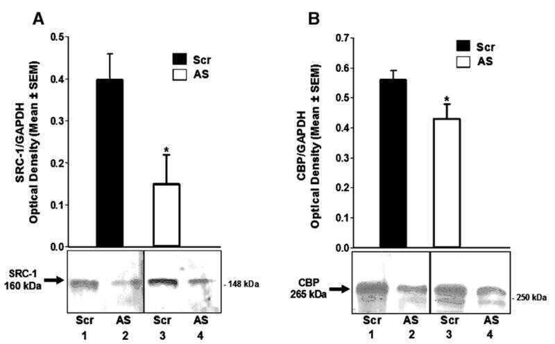
- PA1-847 detects cyclic AMP-responsive enhancer binding protein (CREB) binding protein (CBP) from human and mouse tissues and cells. PA1-847 has been successfully used in ChIP, ICC/IF, Western blot and IHC-P procedures. By Western blot, this antibody detects an ~265 kDa protein representing CBP and a larger, unknown protein at ~400 kDa from HeLa cell lysate. The PA1-847 immunogen is a synthetic peptide corresponding to residues A(162) T S S P A T S Q T G P G I C(176) in the nuclear factor binding domain of human CBP. The PA1-847 immunizing peptide (Cat. # PEP-052) is available for use in neutralization and control experiments.
- Reactivity
- Human, Mouse
- Host
- Rabbit
- Isotype
- IgG
- Vial size
- 100 μg
- Concentration
- 1 mg/mL
- Storage
- -20°C, Avoid Freeze/Thaw Cycles
Submitted references Purine-Induced IFN-γ Promotes Uric Acid Production by Upregulating Xanthine Oxidoreductase Expression.
Convergent organization of aberrant MYB complex controls oncogenic gene expression in acute myeloid leukemia.
Aberrant activation of CYR61 enhancers in colorectal cancer development.
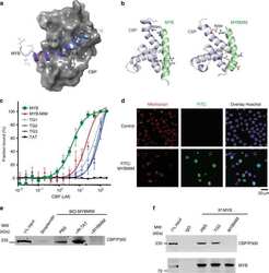
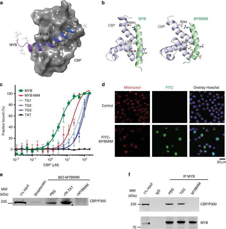
Peptidomimetic blockade of MYB in acute myeloid leukemia.
Expression of glial CBP in steroid mediated neuroprotection in male and female zebra finches.
Research resource: identification of novel coregulators specific for thyroid hormone receptor-β2.
Cells in behaviourally relevant brain regions coexpress nuclear receptor coactivators and ovarian steroid receptors.
Nuclear receptor coactivators function in estrogen receptor- and progestin receptor-dependent aspects of sexual behavior in female rats.
Nuclear receptor coactivators modulate hormone-dependent gene expression in brain and female reproductive behavior in rats.
Role of CBP/P300 in nuclear receptor signalling.
The transcriptional coactivators p300 and CBP are histone acetyltransferases.
The transcriptional coactivators p300 and CBP are histone acetyltransferases.
Wang H, Xie L, Song X, Wang J, Li X, Lin Z, Su T, Liang B, Huang D
Frontiers in immunology 2022;13:773001
Frontiers in immunology 2022;13:773001
Convergent organization of aberrant MYB complex controls oncogenic gene expression in acute myeloid leukemia.
Takao S, Forbes L, Uni M, Cheng S, Pineda JMB, Tarumoto Y, Cifani P, Minuesa G, Chen C, Kharas MG, Bradley RK, Vakoc CR, Koche RP, Kentsis A
eLife 2021 Feb 2;10
eLife 2021 Feb 2;10
Aberrant activation of CYR61 enhancers in colorectal cancer development.
Xie L, Song X, Lin H, Chen Z, Li Q, Guo T, Xu T, Su T, Xu M, Chang X, Wang LK, Liang B, Huang D
Journal of experimental & clinical cancer research : CR 2019 May 22;38(1):213
Journal of experimental & clinical cancer research : CR 2019 May 22;38(1):213
Peptidomimetic blockade of MYB in acute myeloid leukemia.
Ramaswamy K, Forbes L, Minuesa G, Gindin T, Brown F, Kharas MG, Krivtsov AV, Armstrong SA, Still E, de Stanchina E, Knoechel B, Koche R, Kentsis A
Nature communications 2018 Jan 9;9(1):110
Nature communications 2018 Jan 9;9(1):110
Expression of glial CBP in steroid mediated neuroprotection in male and female zebra finches.
Klores M, Moon JT, Duncan KA
Journal of chemical neuroanatomy 2017 Jan;79:32-37
Journal of chemical neuroanatomy 2017 Jan;79:32-37
Research resource: identification of novel coregulators specific for thyroid hormone receptor-β2.
Hahm JB, Privalsky ML
Molecular endocrinology (Baltimore, Md.) 2013 May;27(5):840-59
Molecular endocrinology (Baltimore, Md.) 2013 May;27(5):840-59
Cells in behaviourally relevant brain regions coexpress nuclear receptor coactivators and ovarian steroid receptors.
Tetel MJ, Siegal NK, Murphy SD
Journal of neuroendocrinology 2007 Apr;19(4):262-71
Journal of neuroendocrinology 2007 Apr;19(4):262-71
Nuclear receptor coactivators function in estrogen receptor- and progestin receptor-dependent aspects of sexual behavior in female rats.
Molenda-Figueira HA, Williams CA, Griffin AL, Rutledge EM, Blaustein JD, Tetel MJ
Hormones and behavior 2006 Sep;50(3):383-92
Hormones and behavior 2006 Sep;50(3):383-92
Nuclear receptor coactivators modulate hormone-dependent gene expression in brain and female reproductive behavior in rats.
Molenda HA, Griffin AL, Auger AP, McCarthy MM, Tetel MJ
Endocrinology 2002 Feb;143(2):436-44
Endocrinology 2002 Feb;143(2):436-44
Role of CBP/P300 in nuclear receptor signalling.
Chakravarti D, LaMorte VJ, Nelson MC, Nakajima T, Schulman IG, Juguilon H, Montminy M, Evans RM
Nature 1996 Sep 5;383(6595):99-103
Nature 1996 Sep 5;383(6595):99-103
The transcriptional coactivators p300 and CBP are histone acetyltransferases.
Ogryzko VV, Schiltz RL, Russanova V, Howard BH, Nakatani Y
Cell 1996 Nov 29;87(5):953-9
Cell 1996 Nov 29;87(5):953-9
The transcriptional coactivators p300 and CBP are histone acetyltransferases.
Ogryzko VV, Schiltz RL, Russanova V, Howard BH, Nakatani Y
Cell 1996 Nov 29;87(5):953-9
Cell 1996 Nov 29;87(5):953-9
No comments: Submit comment
Supportive validation
- Submitted by
- Invitrogen Antibodies (provider)
- Main image
- Experimental details
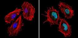
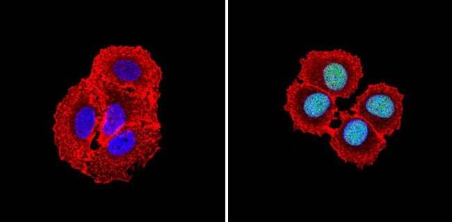
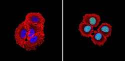
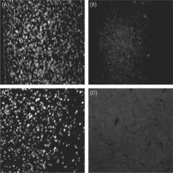
- Immunofluorescent analysis of CREB Binding Protein (green) showing staining in the nucleus of Hela cells (right) compared to a negative control without primary antibody (left). Formalin-fixed cells were permeabilized with 0.1% Triton X-100 in TBS for 5-10 minutes and blocked with 3% BSA-PBS for 30 minutes at room temperature. Cells were probed with a CREB Binding Protein polyclonal antibody (Product # PA1-847) in 3% BSA-PBS at a dilution of 1:100 and incubated overnight at 4ºC in a humidified chamber. Cells were washed with PBST and incubated with a DyLight-conjugated secondary antibody in PBS at room temperature in the dark. Actin was stained using Alexa Fluor 554 (red) and nuclei were stained with Hoechst or DAPI (blue). Images were taken at a magnification of 60x.
- Submitted by
- Invitrogen Antibodies (provider)
- Main image
- Experimental details
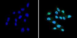
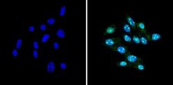
- Immunofluorescent analysis of CREB Binding Protein (green) showing staining in the nucleus of NIH-3T3 cells (right) compared to a negative control without primary antibody (left). Formalin-fixed cells were permeabilized with 0.1% Triton X-100 in TBS for 5-10 minutes and blocked with 3% BSA-PBS for 30 minutes at room temperature. Cells were probed with a CREB Binding Protein polyclonal antibody (Product # PA1-847) in 3% BSA-PBS at a dilution of 1:100 and incubated overnight at 4ºC in a humidified chamber. Cells were washed with PBST and incubated with a DyLight-conjugated secondary antibody in PBS at room temperature in the dark. Nuclei were stained with Hoechst or DAPI (blue). Images were taken at a magnification of 60x.

- Submitted by
- Invitrogen Antibodies (provider)
- Main image

- Experimental details
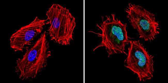
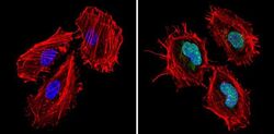
- Immunofluorescent analysis of CREB Binding Protein (green) showing staining in the nucleus of MCF-7 cells (right) compared to a negative control without primary antibody (left). Formalin-fixed cells were permeabilized with 0.1% Triton X-100 in TBS for 5-10 minutes and blocked with 3% BSA-PBS for 30 minutes at room temperature. Cells were probed with a CREB Binding Protein polyclonal antibody (Product # PA1-847) in 3% BSA-PBS at a dilution of 1:100 and incubated overnight at 4ºC in a humidified chamber. Cells were washed with PBST and incubated with a DyLight-conjugated secondary antibody in PBS at room temperature in the dark. Actin was stained using Alexa Fluor 554 (red) and nuclei were stained with Hoechst or DAPI (blue). Images were taken at a magnification of 60x.
- Submitted by
- Invitrogen Antibodies (provider)
- Main image
- Experimental details
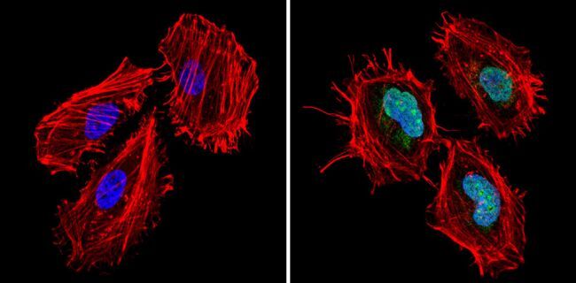
- Immunofluorescent analysis of CREB Binding Protein (green) showing staining in the nucleus of NIH-3T3 cells (right) compared to a negative control without primary antibody (left). Formalin-fixed cells were permeabilized with 0.1% Triton X-100 in TBS for 5-10 minutes and blocked with 3% BSA-PBS for 30 minutes at room temperature. Cells were probed with a CREB Binding Protein polyclonal antibody (Product # PA1-847) in 3% BSA-PBS at a dilution of 1:100 and incubated overnight at 4ºC in a humidified chamber. Cells were washed with PBST and incubated with a DyLight-conjugated secondary antibody in PBS at room temperature in the dark. Nuclei were stained with Hoechst or DAPI (blue). Images were taken at a magnification of 60x.

- Submitted by
- Invitrogen Antibodies (provider)
- Main image
- Experimental details
- Immunofluorescent analysis of CREB Binding Protein (green) showing staining in the nucleus of Hela cells (right) compared to a negative control without primary antibody (left). Formalin-fixed cells were permeabilized with 0.1% Triton X-100 in TBS for 5-10 minutes and blocked with 3% BSA-PBS for 30 minutes at room temperature. Cells were probed with a CREB Binding Protein polyclonal antibody (Product # PA1-847) in 3% BSA-PBS at a dilution of 1:100 and incubated overnight at 4ºC in a humidified chamber. Cells were washed with PBST and incubated with a DyLight-conjugated secondary antibody in PBS at room temperature in the dark. Actin was stained using Alexa Fluor 554 (red) and nuclei were stained with Hoechst or DAPI (blue). Images were taken at a magnification of 60x.
- Submitted by
- Invitrogen Antibodies (provider)
- Main image
- Experimental details
- Immunofluorescent analysis of CREB Binding Protein (green) showing staining in the nucleus of MCF-7 cells (right) compared to a negative control without primary antibody (left). Formalin-fixed cells were permeabilized with 0.1% Triton X-100 in TBS for 5-10 minutes and blocked with 3% BSA-PBS for 30 minutes at room temperature. Cells were probed with a CREB Binding Protein polyclonal antibody (Product # PA1-847) in 3% BSA-PBS at a dilution of 1:100 and incubated overnight at 4ºC in a humidified chamber. Cells were washed with PBST and incubated with a DyLight-conjugated secondary antibody in PBS at room temperature in the dark. Actin was stained using Alexa Fluor 554 (red) and nuclei were stained with Hoechst or DAPI (blue). Images were taken at a magnification of 60x.

Supportive validation
- Submitted by
- Invitrogen Antibodies (provider)
- Main image
- Experimental details
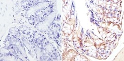
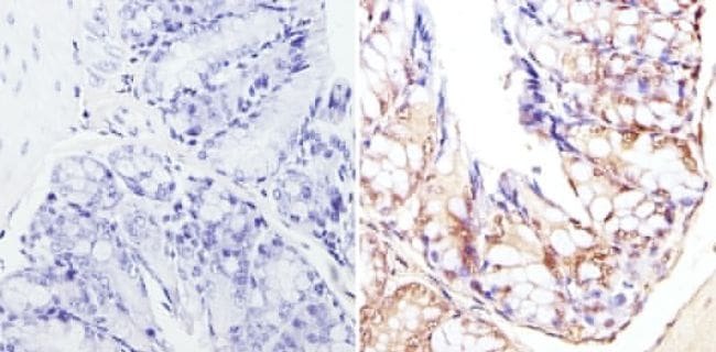
- Immunohistochemistry analysis of CREB Binding Protein showing staining in the cytoplasm and nucleus of paraffin-embedded mouse colon tissue (right) compared to a negative control without primary antibody (left). To expose target proteins, antigen retrieval was performed using 10mM sodium citrate (pH 6.0), microwaved for 8-15 min. Following antigen retrieval, tissues were blocked in 3% H2O2-methanol for 15 min at room temperature, washed with ddH2O and PBS, and then probed with a CREB Binding Protein polyclonal antibody (Product # PA1-847) diluted in 3% BSA-PBS at a dilution of 1:2000 overnight at 4°C in a humidified chamber. Tissues were washed extensively in PBST and detection was performed using an HRP-conjugated secondary antibody followed by colorimetric detection using a DAB kit. Tissues were counterstained with hematoxylin and dehydrated with ethanol and xylene to prep for mounting.
Supportive validation
- Submitted by
- Invitrogen Antibodies (provider)
- Main image
- Experimental details
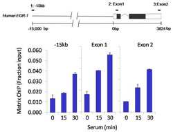
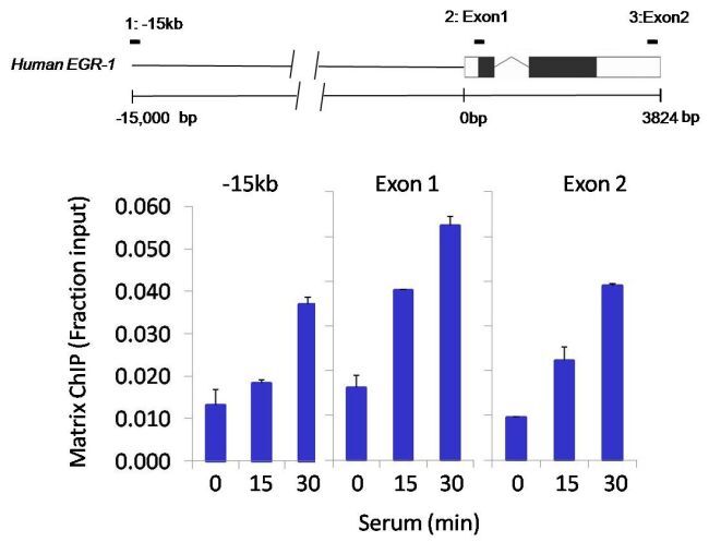
- Chromatin immunoprecipitation analysis of CREB Binding Protein was performed using cross-linked chromatin from 1x10^6 HCT116 colon carcinoma cells treated with serum for 0, 15, and 30 minutes. Immunoprecipitation was performed using a multiplex microplate Matrix ChIP assay (see reference for Matrix ChIP protocol: http://www.ncbi.nlm.nih.gov/pubmed/22098709) with 1.0 µL/100 µL well volume of a CREB Binding Protein polyclonal antibody (Product # PA1-847). Chromatin aliquots from ~1x10^5 cells were used per ChIP pull-down. Quantitative PCR data were done in quadruplicate using 1 µL of eluted DNA in 2 µL SYBR real-time PCR reactions containing primers to amplify -15kb upstream of the Egr1 gene or exon-1 or exon-2 of Egr1. PCR calibration curves were generated for each primer pair from a dilution series of sheared total genomic DNA. Quantitation of immunoprecipitated chromatin is presented as signal relative to the total amount of input chromatin. Results represent the mean +/- SEM for three experiments. A schematic representation of the Egr-1 locus is shown above the data where boxes represent exons (black boxes = translated regions, white boxes = untranslated regions), the zigzag line represents an intron, and the straight line represents upstream sequence. Regions amplified by Egr-1 primers are represented by black bars. Data courtesy of the Innovators Program.

- Submitted by
- Invitrogen Antibodies (provider)
- Main image

- Experimental details
- Chromatin immunoprecipitation analysis of CREB Binding Protein was performed using cross-linked chromatin from 1x10^6 HCT116 colon carcinoma cells treated with serum for 0, 15, and 30 minutes. Immunoprecipitation was performed using a multiplex microplate Matrix ChIP assay (see reference for Matrix ChIP protocol: http://www.ncbi.nlm.nih.gov/pubmed/22098709) with 1.0 µL/100 µL well volume of a CREB Binding Protein polyclonal antibody (Product # PA1-847). Chromatin aliquots from ~1x10^5 cells were used per ChIP pull-down. Quantitative PCR data were done in quadruplicate using 1 µL of eluted DNA in 2 µL SYBR real-time PCR reactions containing primers to amplify -15kb upstream of the Egr1 gene or exon-1 or exon-2 of Egr1. PCR calibration curves were generated for each primer pair from a dilution series of sheared total genomic DNA. Quantitation of immunoprecipitated chromatin is presented as signal relative to the total amount of input chromatin. Results represent the mean +/- SEM for three experiments. A schematic representation of the Egr-1 locus is shown above the data where boxes represent exons (black boxes = translated regions, white boxes = untranslated regions), the zigzag line represents an intron, and the straight line represents upstream sequence. Regions amplified by Egr-1 primers are represented by black bars. Data courtesy of the Innovators Program.
Supportive validation
- Submitted by
- Invitrogen Antibodies (provider)
- Main image
- Experimental details
- NULL
- Submitted by
- Invitrogen Antibodies (provider)
- Main image

- Experimental details
- NULL
- Submitted by
- Invitrogen Antibodies (provider)
- Main image
- Experimental details
- NULL

- Submitted by
- Invitrogen Antibodies (provider)
- Main image

- Experimental details
- NULL