Explore
 Validate
 Learn
Learn Immunocytochemistry
Antibody data
- Antibody Data
- Antigen structure
- References [7]
- Comments [0]
- Validations
- Immunocytochemistry [13]
- Immunohistochemistry [1]
- Chromatin Immunoprecipitation [2]
- Other assay [2]
Submit
Validation data
Reference
Comment
Report error
- Product number
- MA5-13634 - Provider product page

- Provider
- Invitrogen Antibodies
- Product name
- CBP Monoclonal Antibody (NM11)
- Antibody type
- Monoclonal
- Antigen
- Purifed from natural sources
- Description
- MA5-13634 targets p300/CBP (CREB-Binding Protein) in ICC/IF and IHC (P) applications and shows reactivity with Human, Mink, mouse, Non-human primate, and Rat samples. The MA5-13634 immunogen is e1A affinity purified p300 protein.
- Reactivity
- Human, Mouse, Rat
- Host
- Mouse
- Isotype
- IgG
- Antibody clone number
- NM11
- Vial size
- 500 μL
- Concentration
- 0.2 mg/mL
- Storage
- 4°C
Submitted references Alprazolam Prompts HIV-1 Transcriptional Reactivation and Enhances CTL Response Through RUNX1 Inhibition and STAT5 Activation.
USP12 translocation maintains interferon antiviral efficacy by inhibiting CBP acetyltransferase activity.
Benzodiazepines Drive Alteration of Chromatin at the Integrated HIV-1 LTR.
Virus-derived platforms for visualizing protein associations inside cells.
KLF2 Is a novel transcriptional regulator of endothelial proinflammatory activation.
Expression of sex steroid receptors and their co-factors in normal and malignant breast tissue: AIB1 is a carcinoma-specific co-activator.
Expression of sex steroid receptors and their co-factors in normal and malignant breast tissue: AIB1 is a carcinoma-specific co-activator.
Lin A, Elbezanti WO, Schirling A, Ahmed A, Van Duyne R, Cocklin S, Klase Z
Frontiers in neurology 2021;12:663793
Frontiers in neurology 2021;12:663793
USP12 translocation maintains interferon antiviral efficacy by inhibiting CBP acetyltransferase activity.
Liu J, Jin L, Chen X, Yuan Y, Zuo Y, Miao Y, Feng Q, Zhang H, Huang F, Guo T, Zhang L, Zhu L, Qian F, Zhu C, Zheng H
PLoS pathogens 2020 Jan;16(1):e1008215
PLoS pathogens 2020 Jan;16(1):e1008215
Benzodiazepines Drive Alteration of Chromatin at the Integrated HIV-1 LTR.
Elbezanti W, Lin A, Schirling A, Jackson A, Marshall M, Duyne RV, Maldarelli F, Sardo L, Klase Z
Viruses 2020 Feb 9;12(2)
Viruses 2020 Feb 9;12(2)
Virus-derived platforms for visualizing protein associations inside cells.
Miller CL, Arnold MM, Broering TJ, Eichwald C, Kim J, Dinoso JB, Nibert ML
Molecular & cellular proteomics : MCP 2007 Jun;6(6):1027-38
Molecular & cellular proteomics : MCP 2007 Jun;6(6):1027-38
KLF2 Is a novel transcriptional regulator of endothelial proinflammatory activation.
SenBanerjee S, Lin Z, Atkins GB, Greif DM, Rao RM, Kumar A, Feinberg MW, Chen Z, Simon DI, Luscinskas FW, Michel TM, Gimbrone MA Jr, García-Cardeña G, Jain MK
The Journal of experimental medicine 2004 May 17;199(10):1305-15
The Journal of experimental medicine 2004 May 17;199(10):1305-15
Expression of sex steroid receptors and their co-factors in normal and malignant breast tissue: AIB1 is a carcinoma-specific co-activator.
Hudelist G, Czerwenka K, Kubista E, Marton E, Pischinger K, Singer CF
Breast cancer research and treatment 2003 Mar;78(2):193-204
Breast cancer research and treatment 2003 Mar;78(2):193-204
No comments: Submit comment
Supportive validation
- Submitted by
- Invitrogen Antibodies (provider)
- Main image
- Experimental details
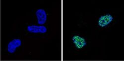
- Immunofluorescent analysis of p300/CBP (CREB-Binding Protein) (green) showing staining in the nucleus of HEK293 cells. Formalin-fixed cells were permeabilized with 0.1% Triton X-100 in TBS for 5-10 minutes and blocked with 3% BSA-PBS for 30 minutes at room temperature. Cells were probed with a p300/CBP (CREB-Binding Protein) monoclonal antibody (Product # MA5-13634) in 3% BSA-PBS at a dilution of 1:100 and incubated overnight at 4 ºC in a humidified chamber. Cells were washed with PBST and incubated with a DyLight-conjugated secondary antibody in PBS at room temperature in the dark. F-actin (red) was stained with a fluorescent red phalloidin and nuclei (blue) were stained with Hoechst or DAPI. Images were taken at a magnification of 60x.

- Submitted by
- Invitrogen Antibodies (provider)
- Main image
- Experimental details
- Immunofluorescent analysis of p300/CBP (CREB-Binding Protein) (green) showing staining in the nucleus of HEK293 cells. Formalin-fixed cells were permeabilized with 0.1% Triton X-100 in TBS for 5-10 minutes and blocked with 3% BSA-PBS for 30 minutes at room temperature. Cells were probed with a p300/CBP (CREB-Binding Protein) monoclonal antibody (Product # MA5-13634) in 3% BSA-PBS at a dilution of 1:100 and incubated overnight at 4 ºC in a humidified chamber. Cells were washed with PBST and incubated with a DyLight-conjugated secondary antibody in PBS at room temperature in the dark. F-actin (red) was stained with a fluorescent red phalloidin and nuclei (blue) were stained with Hoechst or DAPI. Images were taken at a magnification of 60x.
- Submitted by
- Invitrogen Antibodies (provider)
- Main image

- Experimental details
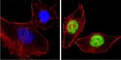
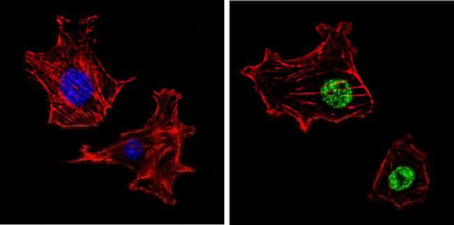
- Immunofluorescent analysis of p300/CBP (CREB-Binding Protein) (green) showing staining in the nucleus of HeLa cells. Formalin-fixed cells were permeabilized with 0.1% Triton X-100 in TBS for 5-10 minutes and blocked with 3% BSA-PBS for 30 minutes at room temperature. Cells were probed with a p300/CBP (CREB-Binding Protein) monoclonal antibody (Product # MA5-13634) in 3% BSA-PBS at a dilution of 1:100 and incubated overnight at 4 ºC in a humidified chamber. Cells were washed with PBST and incubated with a DyLight-conjugated secondary antibody in PBS at room temperature in the dark. F-actin (red) was stained with a fluorescent red phalloidin and nuclei (blue) were stained with Hoechst or DAPI. Images were taken at a magnification of 100x.

- Submitted by
- Invitrogen Antibodies (provider)
- Main image

- Experimental details
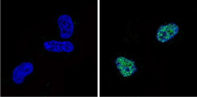
- Immunofluorescent analysis of p300/CBP (CREB-Binding Protein) (green) showing staining in the nucleus of NIH-3T3 cells. Formalin-fixed cells were permeabilized with 0.1% Triton X-100 in TBS for 5-10 minutes and blocked with 3% BSA-PBS for 30 minutes at room temperature. Cells were probed with a p300/CBP (CREB-Binding Protein) monoclonal antibody (Product # MA5-13634) in 3% BSA-PBS at a dilution of 1:100 and incubated overnight at 4 ºC in a humidified chamber. Cells were washed with PBST and incubated with a DyLight-conjugated secondary antibody in PBS at room temperature in the dark. F-actin (red) was stained with a fluorescent red phalloidin and nuclei (blue) were stained with Hoechst or DAPI. Images were taken at a magnification of 60x.
- Submitted by
- Invitrogen Antibodies (provider)
- Main image

- Experimental details
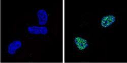
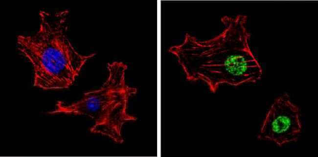
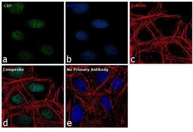
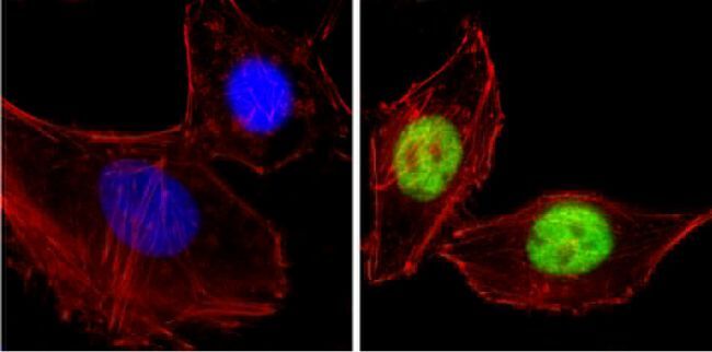
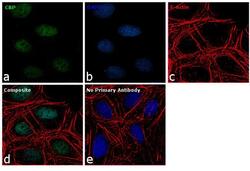
- Immunofluorescence analysis of CBP was performed using 70% confluent log phase U-2 OS cells. The cells were fixed with 4% paraformaldehyde for 10 minutes, permeabilized with 0.1% Triton™ X-100 for 10 minutes, and blocked with 1% BSA for 1 hour at room temperature. The cells were labeled with CBP (NM11) Mouse Monoclonal Antibody (Product # MA5-13634) at 2 µg/mL in 0.1% BSA and incubated for 3 hours at room temperature and then labeled with Goat anti-Mouse IgG (H+L) Superclonal™ Secondary Antibody, Alexa Fluor® 488 conjugate (Product # A28175) at a dilution of 1:2000 for 45 minutes at room temperature (Panel a: green). Nuclei (Panel b: blue) were stained with SlowFade® Gold Antifade Mountant with DAPI (Product # S36938). F-actin (Panel c: red) was stained with Rhodamine Phalloidin (Product # R415, 1:300). Panel d represents the merged image showing nuclear localization. Panel e shows the no primary antibody control. The images were captured at 60X magnification.
- Submitted by
- Invitrogen Antibodies (provider)
- Main image
- Experimental details
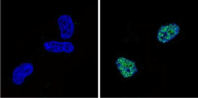
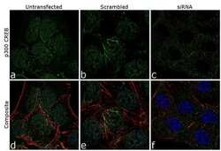
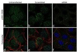
- Knockdown of CBP was achieved by transfecting U-2 OS cells with CBP specific siRNA (Silencer® select Cat # s3497). Immunofluorescence analysis was performed on U-2 OS cells (untransfected, panel a,d), transfected with non-specific scrambled siRNA (panels b,e) and transfected with CBP specific siRNA (panel c,f) Cells were fixed, permeabilized, and labelled with CBP Mouse monoclonal Antibody (Product # MA5-13634, 5 µg/mL), followed by Goat anti-Mouse IgG (H+L) Superclonal™ Secondary Antibody, Alexa Fluor® 488 conjugate (Product # A28175, 1:2000). Nuclei (blue) were stained using SlowFade® Gold Antifade Mountant with DAPI (Product # S36938), and Rhodamine Phalloidin (Product # R415, 1:300) was used for cytoskeletal F-actin (red) staining. Loss of specific signal was observed upon siRNA mediated knockdown (panel c,f) confirming specificity of the antibody to CBP(green). The images were captured at 60X magnification.

- Submitted by
- Invitrogen Antibodies (provider)
- Main image

- Experimental details
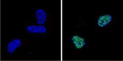
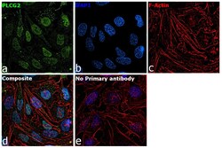
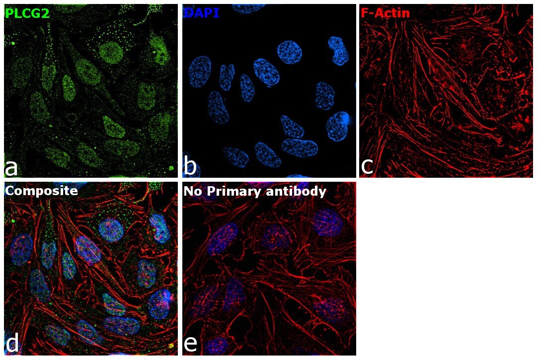
- Immunofluorescence analysis of CBP was performed using 70% confluent log phase HeLa cells. The cells were fixed with 4% paraformaldehyde for 10 minutes, permeabilized with 0.1% Triton™ X-100 for 15 minutes, and blocked with 2% BSA for 45 minutes at room temperature. The cells were labeled with CBP Monoclonal Antibody (NM11) (Product # MA5-13634) at 2 µg/mL in 0.1% BSA, incubated at 4 degree celsius overnight and then labeled with Donkey anti-Mouse IgG (H+L) Highly Cross-Adsorbed Secondary Antibody, Alexa Fluor Plus 488 (Product # A32766), (1:2,000 dilution), for 45 minutes at room temperature (Panel a: Green). Nuclei (Panel b:Blue) were stained with ProLong™ Diamond Antifade Mountant with DAPI (Product # P36962). F-actin (Panel c: Green) was stained with Rhodamine Phalloidin (Product # R415, 1:300). Panel d represents the merged image showing nuclear localization. Panel e represents control cells with no primary antibody to assess background. The images were captured at 60X magnification.

- Submitted by
- Invitrogen Antibodies (provider)
- Main image
- Experimental details
- Immunofluorescent analysis of p300/CBP (CREB-Binding Protein) (green) showing staining in the nucleus of HeLa cells. Formalin-fixed cells were permeabilized with 0.1% Triton X-100 in TBS for 5-10 minutes and blocked with 3% BSA-PBS for 30 minutes at room temperature. Cells were probed with a p300/CBP (CREB-Binding Protein) monoclonal antibody (Product # MA5-13634) in 3% BSA-PBS at a dilution of 1:100 and incubated overnight at 4 ºC in a humidified chamber. Cells were washed with PBST and incubated with a DyLight-conjugated secondary antibody in PBS at room temperature in the dark. F-actin (red) was stained with a fluorescent red phalloidin and nuclei (blue) were stained with Hoechst or DAPI. Images were taken at a magnification of 100x.
- Submitted by
- Invitrogen Antibodies (provider)
- Main image
- Experimental details
- Immunofluorescent analysis of p300/CBP (CREB-Binding Protein) (green) showing staining in the nucleus of HEK293 cells. Formalin-fixed cells were permeabilized with 0.1% Triton X-100 in TBS for 5-10 minutes and blocked with 3% BSA-PBS for 30 minutes at room temperature. Cells were probed with a p300/CBP (CREB-Binding Protein) monoclonal antibody (Product # MA5-13634) in 3% BSA-PBS at a dilution of 1:100 and incubated overnight at 4 ºC in a humidified chamber. Cells were washed with PBST and incubated with a DyLight-conjugated secondary antibody in PBS at room temperature in the dark. F-actin (red) was stained with a fluorescent red phalloidin and nuclei (blue) were stained with Hoechst or DAPI. Images were taken at a magnification of 60x.
- Submitted by
- Invitrogen Antibodies (provider)
- Main image

- Experimental details
- Immunofluorescent analysis of p300/CBP (CREB-Binding Protein) (green) showing staining in the nucleus of NIH-3T3 cells. Formalin-fixed cells were permeabilized with 0.1% Triton X-100 in TBS for 5-10 minutes and blocked with 3% BSA-PBS for 30 minutes at room temperature. Cells were probed with a p300/CBP (CREB-Binding Protein) monoclonal antibody (Product # MA5-13634) in 3% BSA-PBS at a dilution of 1:100 and incubated overnight at 4 ºC in a humidified chamber. Cells were washed with PBST and incubated with a DyLight-conjugated secondary antibody in PBS at room temperature in the dark. F-actin (red) was stained with a fluorescent red phalloidin and nuclei (blue) were stained with Hoechst or DAPI. Images were taken at a magnification of 60x.
- Submitted by
- Invitrogen Antibodies (provider)
- Main image
- Experimental details
- Knockdown of CBP was achieved by transfecting U-2 OS cells with CBP specific siRNA (Silencer® select Cat # s3497). Immunofluorescence analysis was performed on U-2 OS cells (untransfected, panel a,d), transfected with non-specific scrambled siRNA (panels b,e) and transfected with CBP specific siRNA (panel c,f) Cells were fixed, permeabilized, and labelled with CBP Mouse monoclonal Antibody (Product # MA5-13634, 5 µg/mL), followed by Goat anti-Mouse IgG (H+L) Superclonal™ Secondary Antibody, Alexa Fluor® 488 conjugate (Product # A28175, 1:2000). Nuclei (blue) were stained using SlowFade® Gold Antifade Mountant with DAPI (Product # S36938), and Rhodamine Phalloidin (Product # R415, 1:300) was used for cytoskeletal F-actin (red) staining. Loss of specific signal was observed upon siRNA mediated knockdown (panel c,f) confirming specificity of the antibody to CBP(green). The images were captured at 60X magnification.

- Submitted by
- Invitrogen Antibodies (provider)
- Main image
- Experimental details
- Immunofluorescence analysis of CBP was performed using 70% confluent log phase U-2 OS cells. The cells were fixed with 4% paraformaldehyde for 10 minutes, permeabilized with 0.1% Triton™ X-100 for 10 minutes, and blocked with 1% BSA for 1 hour at room temperature. The cells were labeled with CBP (NM11) Mouse Monoclonal Antibody (Product # MA5-13634) at 2 µg/mL in 0.1% BSA and incubated for 3 hours at room temperature and then labeled with Goat anti-Mouse IgG (H+L) Superclonal™ Secondary Antibody, Alexa Fluor® 488 conjugate (Product # A28175) at a dilution of 1:2000 for 45 minutes at room temperature (Panel a: green). Nuclei (Panel b: blue) were stained with SlowFade® Gold Antifade Mountant with DAPI (Product # S36938). F-actin (Panel c: red) was stained with Rhodamine Phalloidin (Product # R415, 1:300). Panel d represents the merged image showing nuclear localization. Panel e shows the no primary antibody control. The images were captured at 60X magnification.

- Submitted by
- Invitrogen Antibodies (provider)
- Main image
- Experimental details
- Immunofluorescence analysis of CBP was performed using 70% confluent log phase HeLa cells. The cells were fixed with 4% paraformaldehyde for 10 minutes, permeabilized with 0.1% Triton™ X-100 for 15 minutes, and blocked with 2% BSA for 45 minutes at room temperature. The cells were labeled with CBP Monoclonal Antibody (NM11) (Product # MA5-13634) at 2 µg/mL in 0.1% BSA, incubated at 4 degree celsius overnight and then labeled with Donkey anti-Mouse IgG (H+L) Highly Cross-Adsorbed Secondary Antibody, Alexa Fluor Plus 488 (Product # A32766), (1:2,000 dilution), for 45 minutes at room temperature (Panel a: Green). Nuclei (Panel b:Blue) were stained with ProLong™ Diamond Antifade Mountant with DAPI (Product # P36962). F-actin (Panel c: Green) was stained with Rhodamine Phalloidin (Product # R415, 1:300). Panel d represents the merged image showing nuclear localization. Panel e represents control cells with no primary antibody to assess background. The images were captured at 60X magnification.
Supportive validation
- Submitted by
- Invitrogen Antibodies (provider)
- Main image

- Experimental details
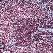
- Formalin-fixed, paraffin-embedded rat spleen stained with p300 antibody using peroxidase-conjugate and AEC chromogen. Note nuclear staining of proliferating cells.
Supportive validation
- Submitted by
- Invitrogen Antibodies (provider)
- Main image
- Experimental details
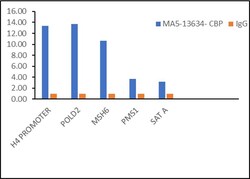
- Chromatin Immunoprecipitation (ChIP) of CBP protein was performed using CBP Monoclonal Antibody (NM11) (Product # MA5-13634, 5 µg) on sheared chromatin from HeLa cells using the MAGnify ChIP System kit (Product # 49-2024). Normal Mouse IgG was used as a negative IP control. The purified DNA was analyzed by qPCR using primers binding to H4 promoter, POLD2, MSH6, PMS1, SAT2 satellite repeats (Inactive). Data is presented as fold enrichment of the antibody signal versus the negative control IgG using the comparative CT method.

- Submitted by
- Invitrogen Antibodies (provider)
- Main image

- Experimental details
- Chromatin Immunoprecipitation (ChIP) of CBP protein was performed using CBP Monoclonal Antibody (NM11) (Product # MA5-13634, 5 µg) on sheared chromatin from HeLa cells using the MAGnify ChIP System kit (Product # 49-2024). Normal Mouse IgG was used as a negative IP control. The purified DNA was analyzed by qPCR using primers binding to H4 promoter, POLD2, MSH6, PMS1, SAT2 satellite repeats (Inactive). Data is presented as fold enrichment of the antibody signal versus the negative control IgG using the comparative CT method.

Supportive validation
- Submitted by
- Invitrogen Antibodies (provider)
- Main image
- Experimental details
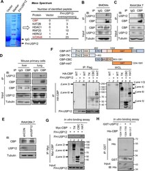
- 10.1371/journal.ppat.1008215.g001 Fig 1 USP12 interacts with the HAT domain of CBP. (A) HEK293T cells were transfected with empty vectors or Flag-HA-USP12 (FH-USP12). The whole cell lysates (WCL) were subjected to immunoprecipitation using Flag (M2) beads. The interacting proteins were analyzed by mass spectrometry (N = 2). (B, C) Immunoprecipitation analysis of the interaction between endogenous USP12 and CBP in BMDMs (B) and RAW264.7 cells (C). (D) Immunoprecipitation analysis of the interaction between endogenous USP12 and CBP in mouse primary lung and liver cells. (E) Western blot analysis of endogenous CBP levels in RAW264.7 cells transfected with either control shRNAs (shCON) or shRNAs against USP12 (shUSP12). (F) The interaction analysis in HEK293T cells cotransfected with FH-USP12 and HA-CBP wild-type (WT) or Myc-CBP mutants including CBP-TK (1-952), CBP-CBC (953-1281), CBP-HAT (1204-1881) as indicated. (G) In vitro binding assay for analysis of the interaction between FH-USP12 and Myc-CBP mutants, including CBP-TK (1-952), CBP-CBC (953-1281), CBP-HAT (1204-1881), which were immunoprecipitated from HEK293T cells transfected with the corresponding plasmids. (H) In vitro binding assay for analysis of the interaction between bacterially expressed GST-USP12 proteins and His-CBP mutant proteins, including CBP-HAT (1324-1700) and CBP-CBC (989-1317), which were purified from E. coli.

- Submitted by
- Invitrogen Antibodies (provider)
- Main image
- Experimental details
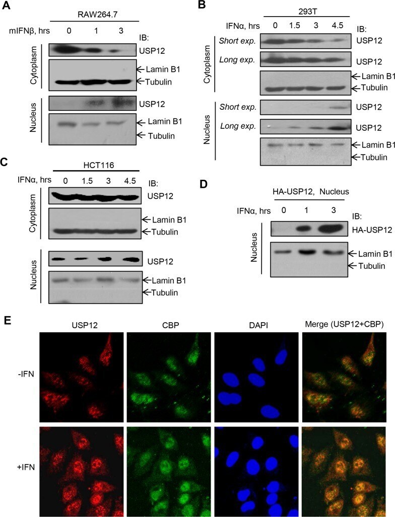
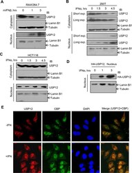
- Fig 4 USP12 translocates from cytoplasm to nucleus in IFN-I signaling. (A) Western blot analysis of USP12 protein levels in the cytoplasm and nucleus of RAW264.7 cells treated with mouse mIFNbeta (500 IU/ml) for 0, 1 and 3 hrs. (B and C) Western blot analysis of USP12 protein levels in the cytoplasm and nucleus of HEK293T (B) or HCT116 (C) cells treated with IFNalpha (1,000 IU/ml) for 0, 1.5, 3 and 4.5 hrs. (D) Western blot analysis of HA-USP12 levels in the nucleus of HEK293T cells transfected with HA-USP12 and then treated with IFNalpha (1,000 IU/ml) as indicated. (E) HeLa cells were treated with IFNalpha (3,000 IU/ml) for 6 hrs. Cellular CBP and USP12 proteins were stained by specific antibodies, and cell nuclei were stained by DAPI. The fluorescent images were captured with the Nikon A1 confocal microscope.