Explore
 Validate
 Learn
Learn Western blot
Antibody data
- Antibody Data
- Antigen structure
- References [5]
- Comments [0]
- Validations
- Western blot [2]
- Immunocytochemistry [8]
- Immunoprecipitation [1]
- Immunohistochemistry [11]
- Chromatin Immunoprecipitation [4]
- Other assay [5]
Submit
Validation data
Reference
Comment
Report error
- Product number
- PA5-27369 - Provider product page

- Provider
- Invitrogen Antibodies
- Product name
- CBP Polyclonal Antibody
- Antibody type
- Polyclonal
- Antigen
- Recombinant full-length protein
- Description
- Recommended positive controls: 293T, A431, HeLa, HepG2, A549, H1299, HCT-116, Vero E6. Predicted reactivity: Mouse (94%), Rat (94%), Rhesus Monkey (100%), Bovine (93%). Store product as a concentrated solution. Centrifuge briefly prior to opening the vial.
- Reactivity
- Human, Mouse
- Host
- Rabbit
- Isotype
- IgG
- Vial size
- 100 μL
- Concentration
- 0.82 mg/mL
- Storage
- Store at 4°C short term. For long term storage, store at -20°C, avoiding freeze/thaw cycles.
Submitted references PER2 mediates CREB-dependent light induction of the clock gene Per1.
Pharmacological targeting of Sam68 functions in colorectal cancer stem cells.
Human T-Cell Lymphotropic Virus Type 1 Transactivator Tax Exploits the XPB Subunit of TFIIH during Viral Transcription.
Tumor-Associated Macrophages Induce Endocrine Therapy Resistance in ER+ Breast Cancer Cells.
DYRK1A interacts with histone acetyl transferase p300 and CBP and localizes to enhancers.
Brenna A, Ripperger JA, Saro G, Glauser DA, Yang Z, Albrecht U
Scientific reports 2021 Nov 5;11(1):21766
Scientific reports 2021 Nov 5;11(1):21766
Pharmacological targeting of Sam68 functions in colorectal cancer stem cells.
Masibag AN, Bergin CJ, Haebe JR, Zouggar A, Shah MS, Sandouka T, Mendes da Silva A, Desrochers FM, Fournier-Morin A, Benoit YD
iScience 2021 Dec 17;24(12):103442
iScience 2021 Dec 17;24(12):103442
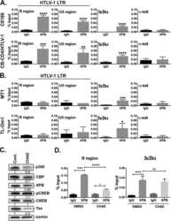
Human T-Cell Lymphotropic Virus Type 1 Transactivator Tax Exploits the XPB Subunit of TFIIH during Viral Transcription.
Martella C, Tollenaere AI, Waast L, Lacombe B, Groussaud D, Margottin-Goguet F, Ramirez BC, Pique C
Journal of virology 2020 Mar 31;94(8)
Journal of virology 2020 Mar 31;94(8)
Tumor-Associated Macrophages Induce Endocrine Therapy Resistance in ER+ Breast Cancer Cells.
Castellaro AM, Rodriguez-Baili MC, Di Tada CE, Gil GA
Cancers 2019 Feb 6;11(2)
Cancers 2019 Feb 6;11(2)
DYRK1A interacts with histone acetyl transferase p300 and CBP and localizes to enhancers.
Li S, Xu C, Fu Y, Lei PJ, Yao Y, Yang W, Zhang Y, Washburn MP, Florens L, Jaiswal M, Wu M, Mohan M
Nucleic acids research 2018 Nov 30;46(21):11202-11213
Nucleic acids research 2018 Nov 30;46(21):11202-11213
No comments: Submit comment
Supportive validation
- Submitted by
- Invitrogen Antibodies (provider)
- Main image
- Experimental details
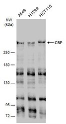
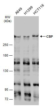
- Western Blot using CBP Polyclonal Antibody (Product # PA5-27369). Various whole cell extracts (30 µg) were separated by 5% SDS-PAGE, and the membrane was blotted with CBP Polyclonal Antibody (Product # PA5-27369) diluted at 1:1,000. The HRP-conjugated anti-rabbit IgG antibody was used to detect the primary antibody.
- Submitted by
- Invitrogen Antibodies (provider)
- Main image

- Experimental details
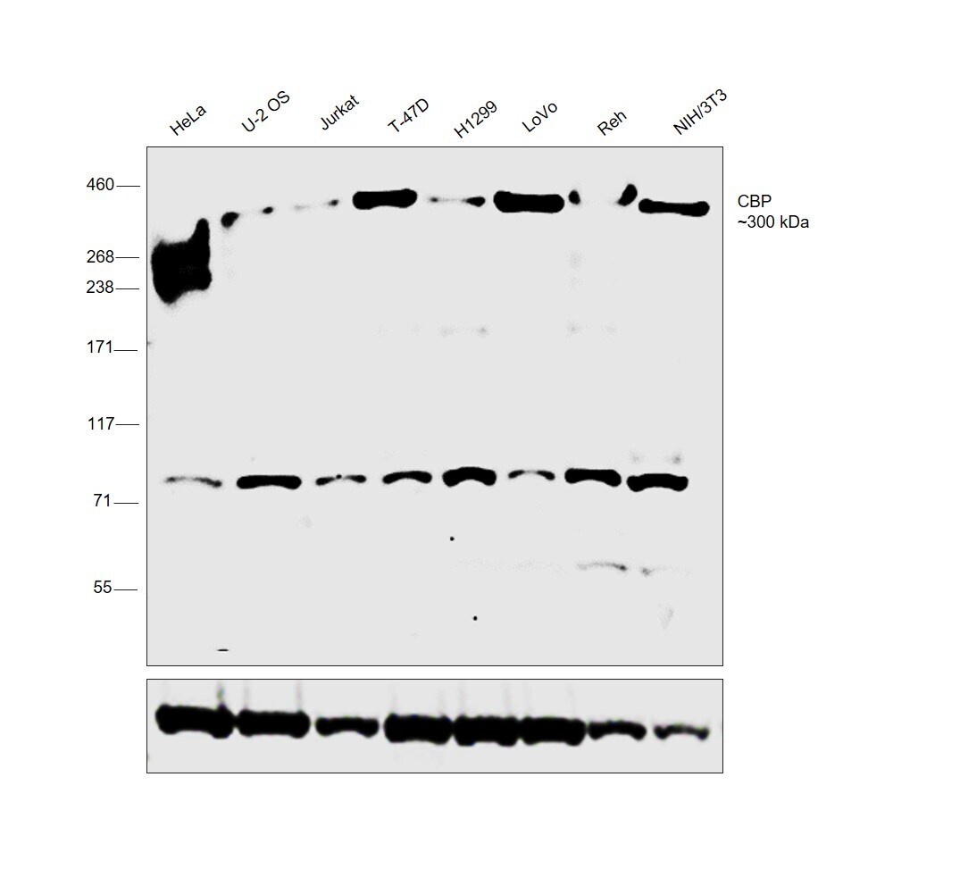
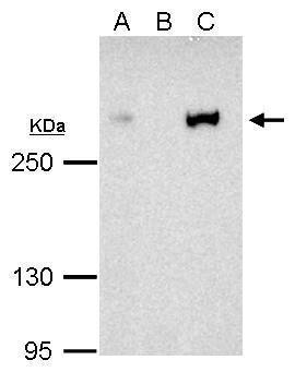
- Western blot was performed using Anti-CBP Polyclonal Antibody (Product # PA5-27369) and a 300kDa band corresponding to CBP was observed across cell lines tested. Nuclear enriched extracts (30 µg lysate) of HeLa (Lane 1), U-2 OS (Lane 2), Jurkat (Lane 3), T-47D (Lane 4), H1299 (Lane 5), LoVo (Lane 6), Reh (Lane 7), NIH/3T3 (Lane 8) were electrophoresed using NuPAGE™ 3-8% Tris-Acetate Protein Gel (Product # EA0378BOX). Resolved proteins were then transferred onto a nitrocellulose membrane (Product # IB23001) by iBlot® 2 Dry Blotting System (Product # IB21001). The blot was probed with the primary antibody (1:1000 dilution) and detected by chemiluminescence with Goat anti-Rabbit IgG (Heavy Chain) Superclonal™ Recombinant Secondary Antibody, HRP (Product # A27036,1:20000 dilution) using the iBright™ FL1500 Imaging System (Product # A44115). Chemiluminescent detection was performed using SuperSignal™ West Pico PLUS Chemiluminescent Substrate (Product # 34580).
Supportive validation
- Submitted by
- Invitrogen Antibodies (provider)
- Main image
- Experimental details
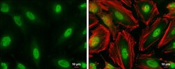
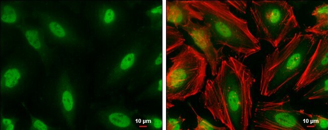
- Immunocytochemistry-Immunofluorescence analysis of CBP was performed in HeLa cells fixed in 4% paraformaldehyde at RT for 15 min. Green: CBP Polyclonal Antibody (Product # PA5-27369) diluted at 1:1000. Red: Phalloidin, a cytoskeleton marker. Scale bar = 10 µm.
- Submitted by
- Invitrogen Antibodies (provider)
- Main image

- Experimental details
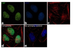
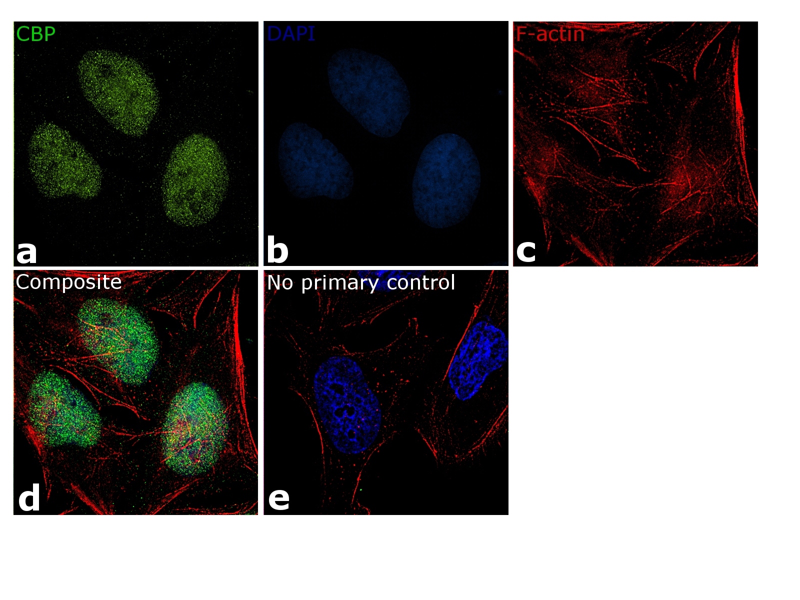
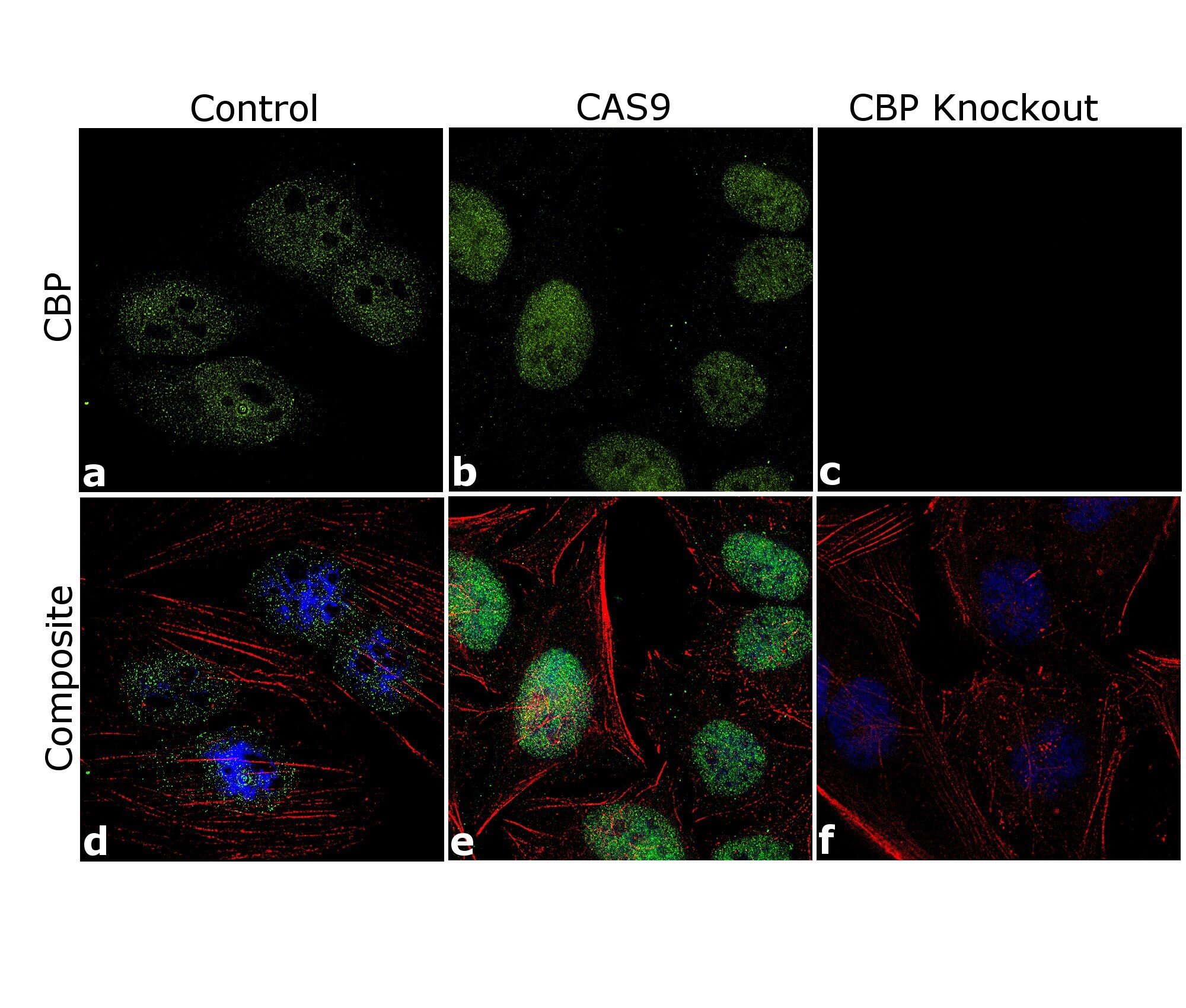
- Immunofluorescence analysis of CBP was done on 70% confluent log phase HeLa cells. The cells were fixed with 4% paraformaldehyde for 15 minutes, permeabilized with 0.1% Triton™ X-100 for 10 minutes, and blocked with 1% BSA for 1 hour at room temperature. The cells were subsequently labeled with CBP Polyclonal Antibody (Product # PA5-27369) at 5µg/mL in 0.1% BSA and incubated for 3 hours at room temperature and then labeled with Goat anti-Rabbit IgG (H+L) Superclonal™ Secondary Antibody, Alexa Fluor® 488 conjugate (Product # A28175) at a dilution of 1:2000 for 45 minutes at room temperature (Panels a-c). Nuclei (Blue) were stained with SlowFade® Gold Antifade Mountant DAPI (Product # S36938). F-actin (Red) was stained with Rhodamine Phalloidin (Product # R415, 1:300). CBP signal was observed in control and scrambled cell lines (panels d and e) and not in the CBP knockout cell line (panel f). The images were captured at 60X magnification.

- Submitted by
- Invitrogen Antibodies (provider)
- Main image
- Experimental details
- Immunofluorescence analysis of CBP was performed using 70% confluent log phase HeLa cells. The cells were fixed with 4% paraformaldehyde for 10 minutes, permeabilized with 0.1% Triton™ X-100 for 10 minutes, and blocked with 1% BSA for 1 hour at room temperature. The cells were labeled with CBP Polyclonal Antibody (Product # PA5-27369) at 5 µg/mL in 0.1% BSA and incubated overnight at 4 degree and then labeled with Goat anti-Rabbit IgG (H+L) Superclonal™ Secondary Antibody, Alexa Fluor® 488 conjugate (Product # A27034) at a dilution of 1:2000 for 45 minutes at room temperature (Panel a: green). Nuclei (Panel b: blue) were stained with SlowFade® Gold Antifade Mountant with DAPI (Product # S36938). F-actin (Panel c: red) was stained with Rhodamine Phalloidin (Product # R415, 1:300). Panel d represents the merged image showing nuclear localization. Panel e represents control cells with no primary antibody to assess background. The images were captured at 60X magnification.
- Submitted by
- Invitrogen Antibodies (provider)
- Main image
- Experimental details
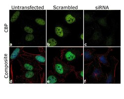
- Knockdown of CBP was achieved by transfecting HeLa cells with CBP specific siRNA (Silencer® select Product # S3497). Immunofluorescence analysis was performed on HeLa cells (untransfected, panel a,d), transfected with non-specific scrambled siRNA (panels b,e) and transfected with CBP specific siRNA (panel c,f). Cells were fixed, permeabilized, and labelled with CBP Polyclonal Antibody (Product # PA5-27369, 5µg/mL), followed by Goat anti-Rabbit IgG (H+L) Superclonal™ Secondary Antibody, Alexa Fluor® 488 conjugate (Product # A27034, 1:2000). Nuclei (blue) were stained using SlowFade® Gold Antifade Mountant with DAPI (Product # S36938), and Rhodamine Phalloidin (Product # R415, 1:300) was used for cytoskeletal F-actin (red) staining. Reduction of specific signal was observed upon siRNA mediated knockdown (panel c,f) confirming specificity of the antibody to CBP (green). The images were captured at 60X magnification.

- Submitted by
- Invitrogen Antibodies (provider)
- Main image

- Experimental details
- Immunofluorescence analysis of CBP was done on 70% confluent log phase HeLa cells. The cells were fixed with 4% paraformaldehyde for 15 minutes, permeabilized with 0.1% Triton™ X-100 for 10 minutes, and blocked with 1% BSA for 1 hour at room temperature. The cells were subsequently labeled with CBP Polyclonal Antibody (Product # PA5-27369) at 5µg/mL in 0.1% BSA and incubated for 3 hours at room temperature and then labeled with Goat anti-Rabbit IgG (H+L) Superclonal™ Secondary Antibody, Alexa Fluor® 488 conjugate (Product # A28175) at a dilution of 1:2000 for 45 minutes at room temperature (Panels a-c). Nuclei (Blue) were stained with SlowFade® Gold Antifade Mountant DAPI (Product # S36938). F-actin (Red) was stained with Rhodamine Phalloidin (Product # R415, 1:300). CBP signal was observed in control and scrambled cell lines (panels d and e) and not in the CBP knockout cell line (panel f). The images were captured at 60X magnification.
- Submitted by
- Invitrogen Antibodies (provider)
- Main image

- Experimental details
- Immunofluorescence analysis of CBP was performed using 70% confluent log phase HeLa cells. The cells were fixed with 4% paraformaldehyde for 10 minutes, permeabilized with 0.1% Triton™ X-100 for 10 minutes, and blocked with 1% BSA for 1 hour at room temperature. The cells were labeled with CBP Polyclonal Antibody (Product # PA5-27369) at 5 µg/mL in 0.1% BSA and incubated overnight at 4 degree and then labeled with Goat anti-Rabbit IgG (Heavy Chain) Superclonal™ Secondary Antibody, Alexa Fluor® 488 conjugate (Product # A27034) at a dilution of 1:2000 for 45 minutes at room temperature (Panel a: green). Nuclei (Panel b: blue) were stained with SlowFade® Gold Antifade Mountant with DAPI (Product # S36938). F-actin (Panel c: red) was stained with Rhodamine Phalloidin (Product # R415, 1:300). Panel d represents the merged image showing nuclear localization. Panel e represents control cells with no primary antibody to assess background. The images were captured at 60X magnification.

- Submitted by
- Invitrogen Antibodies (provider)
- Main image

- Experimental details
- Knockdown of CBP was achieved by transfecting HeLa cells with CBP specific siRNA (Silencer® select Product # S3497). Immunofluorescence analysis was performed on HeLa cells (untransfected, panel a,d), transfected with non-specific scrambled siRNA (panels b,e) and transfected with CBP specific siRNA (panel c,f). Cells were fixed, permeabilized, and labelled with CBP Polyclonal Antibody (Product # PA5-27369, 5µg/mL), followed by Goat anti-Rabbit IgG (Heavy Chain) Superclonal™ Secondary Antibody, Alexa Fluor® 488 conjugate (Product # A27034, 1:2000). Nuclei (blue) were stained using SlowFade® Gold Antifade Mountant with DAPI (Product # S36938), and Rhodamine Phalloidin (Product # R415, 1:300) was used for cytoskeletal F-actin (red) staining. Reduction of specific signal was observed upon siRNA mediated knockdown (panel c,f) confirming specificity of the antibody to CBP (green). The images were captured at 60X magnification.

- Submitted by
- Invitrogen Antibodies (provider)
- Main image

- Experimental details
- Immunocytochemistry-Immunofluorescence analysis of CBP was performed in HeLa cells fixed in 4% paraformaldehyde at RT for 15 min. Green: CBP Polyclonal Antibody (Product # PA5-27369) diluted at 1:1000. Red: Phalloidin, a cytoskeleton marker. Scale bar = 10 µm.

Supportive validation
- Submitted by
- Invitrogen Antibodies (provider)
- Main image

- Experimental details
- CBP antibody immunoprecipitates CBP protein in IP experiments. IP Sample: 1,000 µg HeLa whole cell lysate/extract A. 40 µg HeLa whole cell lysate/extract B. Control with 2.5 µg of preimmune rabbit IgG C. Immunoprecipitation of CBP protein by 2.5 µg of CBP antibody (Product # PA5-27369) 12% SDS-PAGE The immunoprecipitated CBP protein was detected by CBP antibody (Product # PA5-27369) diluted at 1:1,000.
Supportive validation
- Submitted by
- Invitrogen Antibodies (provider)
- Main image
- Experimental details
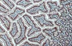
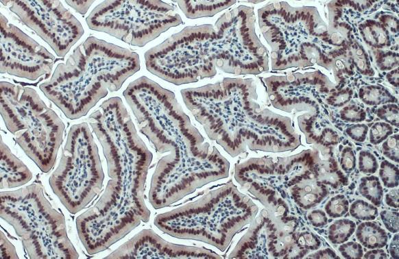
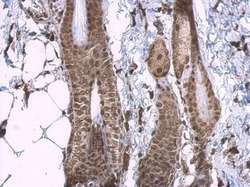
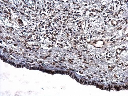
- CBP Polyclonal Antibody detects CBP protein at nucleus by immunohistochemical analysis. Sample: Paraffin-embedded mouse intestine. CBP stained by CBP Polyclonal Antibody (Product # PA5-27369) diluted at 1:500. Antigen Retrieval: Citrate buffer, pH 6.
- Submitted by
- Invitrogen Antibodies (provider)
- Main image
- Experimental details
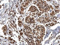
- CBP Polyclonal Antibody detects CBP protein at nucleus on human breast carcinoma by immunohistochemical analysis. Sample: Paraffin-embedded human breast carcinoma. CBP Polyclonal Antibody (Product # PA5-27369) dilution: 1:750. Antigen Retrieval: EDTA based buffer, pH 8.0, 15 min.

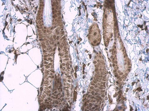
- Submitted by
- Invitrogen Antibodies (provider)
- Main image
- Experimental details
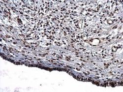
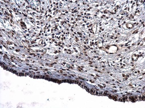
- CBP Polyclonal Antibody detects CBP protein at nucleus on human cervical carcinoma by immunohistochemical analysis. Sample: Paraffin-embedded human cervical carcinoma. CBP Polyclonal Antibody (Product # PA5-27369) dilution: 1:750. Antigen Retrieval: EDTA based buffer, pH 8.0, 15 min.
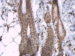
- Submitted by
- Invitrogen Antibodies (provider)
- Main image
- Experimental details
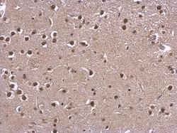
- CBP Polyclonal Antibody detects CBP protein at nucleus on mouse muscle by immunohistochemical analysis. Sample: Paraffin-embedded mouse muscle. CBP Polyclonal Antibody (Product # PA5-27369) dilution: 1:750. Antigen Retrieval: EDTA based buffer, pH 8.0, 15 min.

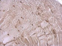
- Submitted by
- Invitrogen Antibodies (provider)
- Main image
- Experimental details
- CBP Polyclonal Antibody detects CBP protein at nucleus on mouse skin by immunohistochemical analysis. Sample: Paraffin-embedded mouse skin. CBP Polyclonal Antibody (Product # PA5-27369) dilution: 1:750. Antigen Retrieval: EDTA based buffer, pH 8.0, 15 min.
- Submitted by
- Invitrogen Antibodies (provider)
- Main image
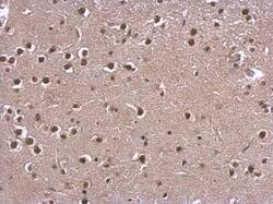
- Experimental details
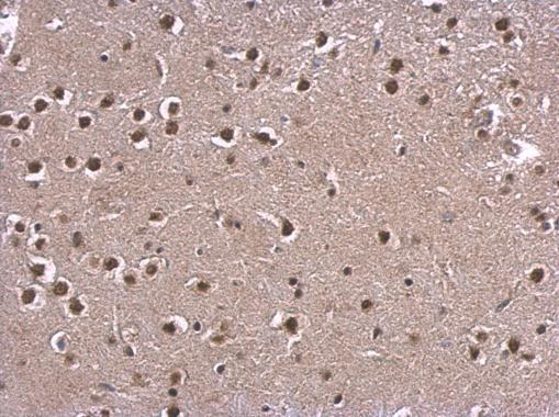
- CBP Polyclonal Antibody detects CBP protein at nucleus on mouse fore brain by immunohistochemical analysis. Sample: Paraffin-embedded mouse fore brain. CBP Polyclonal Antibody (Product # PA5-27369) dilution: 1:750. Antigen Retrieval: EDTA based buffer, pH 8.0, 15 min.
- Submitted by
- Invitrogen Antibodies (provider)
- Main image

- Experimental details
- CBP Polyclonal Antibody detects CBP protein at nucleus by immunohistochemical analysis. Sample: Paraffin-embedded mouse intestine. CBP stained by CBP Polyclonal Antibody (Product # PA5-27369) diluted at 1:500. Antigen Retrieval: Citrate buffer, pH 6.

- Submitted by
- Invitrogen Antibodies (provider)
- Main image

- Experimental details
- CBP Polyclonal Antibody detects CBP protein at nucleus on human breast carcinoma by immunohistochemical analysis. Sample: Paraffin-embedded human breast carcinoma. CBP Polyclonal Antibody (Product # PA5-27369) dilution: 1:750. Antigen Retrieval: EDTA based buffer, pH 8.0, 15 min.

- Submitted by
- Invitrogen Antibodies (provider)
- Main image
- Experimental details
- CBP Polyclonal Antibody detects CBP protein at nucleus on mouse skin by immunohistochemical analysis. Sample: Paraffin-embedded mouse skin. CBP Polyclonal Antibody (Product # PA5-27369) dilution: 1:750. Antigen Retrieval: EDTA based buffer, pH 8.0, 15 min.

- Submitted by
- Invitrogen Antibodies (provider)
- Main image
- Experimental details
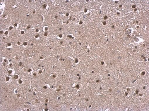
- CBP Polyclonal Antibody detects CBP protein at nucleus on mouse fore brain by immunohistochemical analysis. Sample: Paraffin-embedded mouse fore brain. CBP Polyclonal Antibody (Product # PA5-27369) dilution: 1:750. Antigen Retrieval: EDTA based buffer, pH 8.0, 15 min.
- Submitted by
- Invitrogen Antibodies (provider)
- Main image

- Experimental details
- CBP Polyclonal Antibody detects CBP protein at nucleus on human cervical carcinoma by immunohistochemical analysis. Sample: Paraffin-embedded human cervical carcinoma. CBP Polyclonal Antibody (Product # PA5-27369) dilution: 1:750. Antigen Retrieval: EDTA based buffer, pH 8.0, 15 min.
Supportive validation
- Submitted by
- Invitrogen Antibodies (provider)
- Main image

- Experimental details
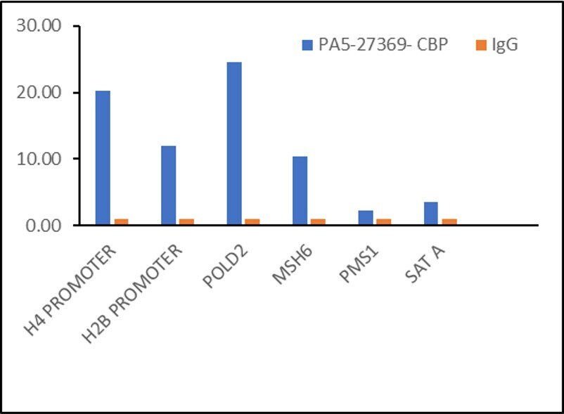
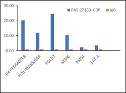
- Chromatin Immunoprecipitation (ChIP) of CBP protein was performed using CBP Polyclonal Antibody (Product # PA5-27369, 5 µg) on sheared chromatin from HeLa cells using the MAGnify ChIP System kit (Product # 49-2024). Normal Rabbit IgG was used as a negative IP control. The purified DNA was analyzed by qPCR using primers binding to H4 promoter, H2B promoter, POLD2, MSH6, PMS1, SAT2 satellite repeats (Inactive). Data is presented as fold enrichment of the antibody signal versus the negative control IgG using the comparative CT method.
- Submitted by
- Invitrogen Antibodies (provider)
- Main image
- Experimental details
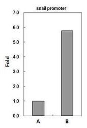
- CREBBP antibody immunoprecipitates CREBBP protein-DNA complex in ChIP experiments. ChIP Sample: HeLa whole cell lysate/extract. A: 5 µg preimmune rabbit IgG. B: 5 µg of CREBBP antibody (Product # PA5-27369). The precipitated DNA was detected by PCR with primer set targeting to snail promoter.
- Submitted by
- Invitrogen Antibodies (provider)
- Main image

- Experimental details
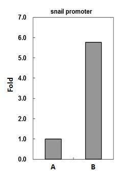
- CREBBP antibody immunoprecipitates CREBBP protein-DNA complex in ChIP experiments. ChIP Sample: HeLa whole cell lysate/extract. A: 5 µg preimmune rabbit IgG. B: 5 µg of CREBBP antibody (Product # PA5-27369). The precipitated DNA was detected by PCR with primer set targeting to snail promoter.

- Submitted by
- Invitrogen Antibodies (provider)
- Main image
- Experimental details
- Chromatin Immunoprecipitation (ChIP) of CBP protein was performed using CBP Polyclonal Antibody (Product # PA5-27369, 5 µg) on sheared chromatin from HeLa cells using the MAGnify ChIP System kit (Product # 49-2024). Normal Rabbit IgG was used as a negative IP control. The purified DNA was analyzed by qPCR using primers binding to H4 promoter, H2B promoter, POLD2, MSH6, PMS1, SAT2 satellite repeats (Inactive). Data is presented as fold enrichment of the antibody signal versus the negative control IgG using the comparative CT method.

Supportive validation
- Submitted by
- Invitrogen Antibodies (provider)
- Main image
- Experimental details
- NULL

- Submitted by
- Invitrogen Antibodies (provider)
- Main image

- Experimental details
- CBP antibody immunoprecipitates CBP protein in IP experiments. IP Sample: 1,000 µg HeLa whole cell lysate/extract A. 40 µg HeLa whole cell lysate/extract B. Control with 2.5 µg of preimmune rabbit IgG C. Immunoprecipitation of CBP protein by 2.5 µg of CBP antibody (Product # PA5-27369) 12% SDS-PAGE The immunoprecipitated CBP protein was detected by CBP antibody (Product # PA5-27369) diluted at 1:1,000.

- Submitted by
- Invitrogen Antibodies (provider)
- Main image
- Experimental details
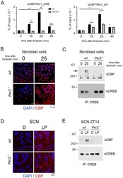
- Figure 4 Modulation of the interaction between CREB and CBP by PER2. ( A ) Left panel: Chromatin immunoprecipitation (ChIP) of CBP on the CRE-element of the Per1 promoter in wt (black bars) and Per2 KO (grey bars) fibroblast cell lines after forskolin treatment. Right panel: Control ChIP on a promoter region of Per1 without a CRE-element . Values are the mean +- SD. Student's t-test with Welch's correction, n = 3, * p < 0.05, ** p < 0.01. Two-way ANOVA indicates a significantly different time profile between the two genotypes, left panel: **** p < 0.0001, right panel: ** p < 0.01. ( B ) Immunohistochemistry (IHC) of wt and Per2 -/- cells before and after forskolin treatment. CBP (red signal) is induced after forskolin treatment in both genotypes. Scale bar: 17 um. ( C ) IP of CREB pulls down CBP in wt but not Per2 -/- cells after forskolin treatment, kD = kilo Dalton. ( D ) IHC of wt and Per2 -/- SCN before and after a light pulse (LP) at ZT14. CBP (red signal) is induced after LP in both genotypes. Scale bar: 7 um. ( E ) IP of CREB pulls down CBP in wt SCN extracts after LP, but not in Per2 -/- SCN extracts. kD = kilo Dalton.

- Submitted by
- Invitrogen Antibodies (provider)
- Main image
- Experimental details
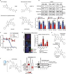
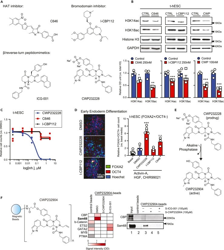
- Figure 1 Reverse/beta-turn peptidomimetic compounds are direct interactors of Sam68 (A) Chemical structure of HAT inhibitor C646 and bromodomain ligand I-CBP112, as well as beta-turn peptidomimetics ICG-001 and CWP232228. (B) Western blot analysis of CBP-catalyzed H3K14ac and H3K18ac histone acetylation marks in C646 (0.25 muM), I-CBP112 (0.25 muM), and CWP232228 (0.1 muM) t-hESCs versus control DMSO. Total histone H3 and GAPDH were used as loading control. Relative OD signal quantification versus H3 intensity is presented (C646: n = 3, I-CBP112: n = 5, CWP232228: n >= 3, *: p = 0.0183, **: p = 0.0078, ***: p
- Submitted by
- Invitrogen Antibodies (provider)
- Main image

- Experimental details
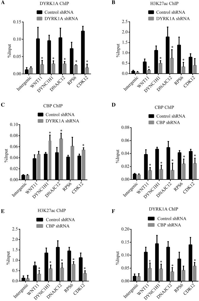
- Figure 6. DYRK1A is required for p300/CBP mediated H3K27 acetylation at its target enhancers ( A - C ) ChIP analysis of DYRK1A, H3K27acetylation and CBP in cells treated with control or DYRK1A shRNA. DYRK1A bound enhancer sites (near DNAJC12, DYNC1H1 and WNT11) and at TSS (RPS6 and CDK12) were probed for enrichment. ( D - F ) ChIP analysis of CBP, H3K27acetylation and DYRK1A in cells treated with control or CBP shRNA. Sites probed as in (A-C). Data represent the mean +- SD ( n = 3 biological replicates). Student's t test were done to compare samples. P value = * ( P < 0.05).