Explore
 Validate
 Learn
Learn Western blot
 Immunocytochemistry
Antibody data
- Antibody Data
- Antigen structure
- References [2]
- Comments [0]
- Validations
- Western blot [2]
Submit
Validation data
Reference
Comment
Report error
- Product number
- MAB2676 - Provider product page

- Provider
- R&D Systems
- Product name
- Human/Mouse/Rat CBP Antibody
- Antibody type
- Monoclonal
- Description
- Protein A or G purified from hybridoma culture supernatant. Detects human, mouse and rat CBP.
- Reactivity
- Human, Mouse, Rat
- Host
- Mouse
- Conjugate
- Unconjugated
- Antigen sequence
Q92793- Isotype
- IgG
- Antibody clone number
- 257003
- Vial size
- 100 ug
- Concentration
- LYOPH
- Storage
- Use a manual defrost freezer and avoid repeated freeze-thaw cycles. 12 months from date of receipt, -20 to -70 °C as supplied. 1 month, 2 to 8 °C under sterile conditions after reconstitution. 6 months, -20 to -70 °C under sterile conditions after reconstitution.
Submitted references Respiratory syncytial virus impairs macrophage IFN-alpha/beta- and IFN-gamma-stimulated transcription by distinct mechanisms.
Effect of cAMP on TGFbeta1-induced corneal keratocyte-myofibroblast transformation.
Senft AP, Taylor RH, Lei W, Campbell SA, Tipper JL, Martinez MJ, Witt TL, Clay CC, Harrod KS
American journal of respiratory cell and molecular biology 2010 Apr;42(4):404-14
American journal of respiratory cell and molecular biology 2010 Apr;42(4):404-14
Effect of cAMP on TGFbeta1-induced corneal keratocyte-myofibroblast transformation.
Xing D, Bonanno JA
Investigative ophthalmology & visual science 2009 Feb;50(2):626-33
Investigative ophthalmology & visual science 2009 Feb;50(2):626-33
No comments: Submit comment
Supportive validation
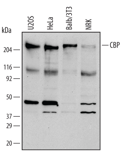
- Submitted by
- R&D Systems (provider)
- Main image
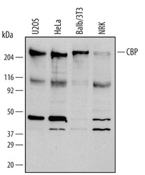
- Experimental details
- Detection of Human/Mouse/Rat CBP by Western Blot. Western blot shows lysates of NRK rat normal kidney cell line, U2OS human osteosarcoma cell line, HeLa human cervical epithelial carcinoma cell line, and Balb/3T3 mouse embryonic fibroblast cell line. PVDF membrane was probed with 0.4 µg/mL of Human/Mouse/Rat CBP Monoclonal Antibody (Catalog # MAB2676) followed by HRP-conjugated Anti-Mouse IgG Secondary Antibody (Catalog # HAF007). A specific band was detected for CBP at approximately 220 kDa (as indicated). This experiment was conducted under reducing conditions and using Immunoblot Buffer Group 1.
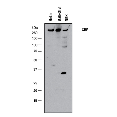
- Submitted by
- R&D Systems (provider)
- Main image
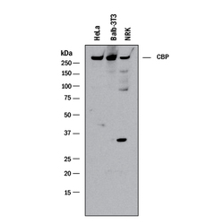
- Experimental details
- Detection of Human, Mouse, and Rat CBP by Western Blot. Western blot shows lysates of HeLa human cervical epithelial carcinoma cell line, Balb/3T3 mouse embryonic fibroblast cell line, and NRK rat normal kidney cell line. PVDF membrane was probed with 0.5 µg/mL of Mouse Anti-Human/Mouse/Rat CBP Monoclonal Antibody (Catalog # MAB2676) followed by HRP-conjugated Anti-Mouse IgG Secondary Antibody (Catalog # HAF018). A specific band was detected for CBP at approximately 265 kDa (as indicated). This experiment was conducted under reducing conditions and using Immunoblot Buffer Group 1.