 Explore
 Validate
 Learn
Learn Western blot
 Immunohistochemistry
Antibody data
- Antibody Data
- Antigen structure
- References [0]
- Comments [0]
- Validations
- Western blot [5]
- Immunocytochemistry [2]
Submit
Validation data
Reference
Comment
Report error
- Product number
- MA5-24791 - Provider product page

- Provider
- Invitrogen Antibodies
- Product name
- EWSR1 Monoclonal Antibody (5H7)
- Antibody type
- Monoclonal
- Antigen
- Purifed from natural sources
- Reactivity
- Human, Mouse, Rat, Canine
- Host
- Mouse
- Isotype
- IgG
- Antibody clone number
- 5H7
- Vial size
- 100 µL
- Concentration
- 1 mg/mL
- Storage
- Store at 4°C short term. For long term storage, store at -20°C, avoiding freeze/thaw cycles.
No comments: Submit comment
Supportive validation
- Submitted by
- Invitrogen Antibodies (provider)
- Main image
- Experimental details
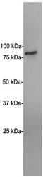
- Western blot analysis of EWSR1 in HeLa cells. The membrane was probed with EWSR1 monoclonal antibody (Product # MA5-24791).

- Submitted by
- Invitrogen Antibodies (provider)
- Main image

- Experimental details
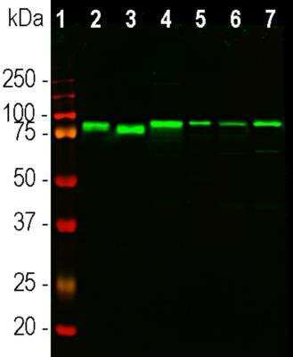
- Western blot analysis of EWSR1 in different cell line lysates. Samples were incubated in EWSR1 monoclonal antibody (Product # MA5-24791 using a dilution of 1:1000. Antibody in green: [1] protein standard (Red), [2] HeLa, [3] HEK293, [4] mouse NIH-3T3, [5] rat PC12, [6] equine NBL6 and [7] canine A72 cells. The strong band at ~80 kDa corresponds to the EWS protein seen in all species tested.
- Submitted by
- Invitrogen Antibodies (provider)
- Main image

- Experimental details
- Western blot analysis of EWSR1 in different cell line lysates. Samples were incubated in EWSR1 monoclonal antibody (Product # MA5-24791 using a dilution of 1:1000. Antibody in green: [1] protein standard (Red), [2] HeLa, [3] HEK293, [4] mouse NIH-3T3, [5] rat PC12, [6] equine NBL6 and [7] canine A72 cells. The strong band at ~80 kDa corresponds to the EWS protein seen in all species tested.

- Submitted by
- Invitrogen Antibodies (provider)
- Main image

- Experimental details
- Western blot analysis of EWSR1 in HEK293T and huh-7 lysate. Samples were incubated in EWSR1 monoclonal antibody (Product # MA5-24791). A specific band for EWSR1 (observed molecular weight ~95 kDa).

- Submitted by
- Invitrogen Antibodies (provider)
- Main image

- Experimental details
- Western blot was performed using anti-EWSR1 Monoclonal Antibody (5H7) (Product # MA5-24791) and 68, 61 kDa bands corresponding to EWSR1 was observed across the cell lines and tissue tested. Modified whole cell extracts (1% SDS) (30 µg lysate) of MOLT-4 (Lane 1), HeLa (Lane 2), Hep G2 (Lane 3), NIH/3T3 (Lane 4), MCF7 (Lane 5), RAW 264.7 (Lane 6), Jurkat (Lane 7) and Rat Testis (Lane 8) were electrophoresed using Novex® NuPAGE™ 4-12% Bis-Tris Protein Gel (Product # NP0322BOX). Resolved proteins were then transferred onto a nitrocellulose membrane (Product # IB23001) by iBlot® 2 Dry Blotting System (Product # IB21001). The blot was probed with the primary antibody (1:1000 dilution) and detected by chemiluminescence with Goat anti-Mouse IgG (H+L), Superclonal™ Recombinant Secondary Antibody, HRP (Product # A28177, 1:4000 dilution) using the iBright FL 1000 (Product # A32752). Chemiluminescent detection was performed using Novex® ECL Chemiluminescent Substrate Reagent Kit (Product # WP20005).

Supportive validation
- Submitted by
- Invitrogen Antibodies (provider)
- Main image
- Experimental details
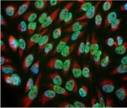
- Immunocytochemistry analysis of EWSR1 in HeLa cells. Samples were incubated in EWSR1 monoclonal antibody (Product # MA5-24791). Antibody (green) and chicken antibody to vimentin. Blue is a DNA stain, and it is clear that in these cells EWSR1 is localized along with the DNA in the nucleus.
- Submitted by
- Invitrogen Antibodies (provider)
- Main image
- Experimental details
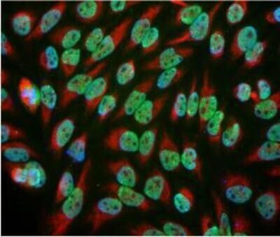
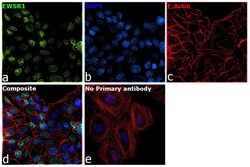
- Immunofluorescence analysis of EWSR1 was performed using 70% confluent log phase MCF7 cells. The cells were fixed with 4% paraformaldehyde for 10 minutes, permeabilized with 0.1% Triton™ X-100 for 15 minutes, and blocked with 2% BSA for 1 hour at room temperature. The cells were labeled with EWSR1 Monoclonal Antibody (5H7) (Product # MA5-24791) at 1:100 dilution in 0.1% BSA, incubated at 4 degree Celsius overnight and then with Goat anti-Mouse IgG (H+L) Cross-Adsorbed Secondary Antibody, Alexa Fluor 488 (Product # A32723) at a dilution of 1:2000 for 45 minutes at room temperature (Panel a: green). Nuclei (Panel b: blue) were stained with ProLong™ Diamond Antifade Mountant with DAPI (Product # P36962). F-actin (Panel c: red) was stained with Rhodamine Phalloidin (Product # R415, 1:300). Panel d represents the merged image showing nuclear localization. Panel e represents control cells with no primary antibody to assess background. The images were captured at 60X magnification.
