Explore
 Validate
 Learn
Learn Western blot
Antibody data
- Antibody Data
- Antigen structure
- References [3]
- Comments [0]
- Validations
- Western blot [1]
- Immunohistochemistry [1]
Submit
Validation data
Reference
Comment
Report error
- Product number
- sc-13121 - Provider product page

- Provider
- Santa Cruz Biotechnology
- Proper citation
- Santa Cruz Biotechnology Cat#sc-13121, RRID:AB_627596
- Product name
- Anti-FGFR3
- Antibody type
- Monoclonal
- Antigen
- Recombinant full-length protein
- Reactivity
- Human
- Host
- Mouse
Submitted references Rapidly acquired resistance to EGFR tyrosine kinase inhibitors in NSCLC cell lines through de-repression of FGFR2 and FGFR3 expression.
Ectopic expression of wild-type FGFR3 cooperates with MYC to accelerate development of B-cell lineage neoplasms.
Increased expression of fibroblast growth factor receptor 3 in CD34+ BCR-ABL+ cells from patients with chronic myeloid leukemia
Ware KE, Marshall ME, Heasley LR, Marek L, Hinz TK, Hercule P, Helfrich BA, Doebele RC, Heasley LE
PloS one 2010 Nov 29;5(11):e14117
PloS one 2010 Nov 29;5(11):e14117
Ectopic expression of wild-type FGFR3 cooperates with MYC to accelerate development of B-cell lineage neoplasms.
Zingone A, Cultraro CM, Shin DM, Bean CM, Morse HC 3rd, Janz S, Kuehl WM
Leukemia : official journal of the Leukemia Society of America, Leukemia Research Fund, U.K 2010 Jun;24(6):1171-8
Leukemia : official journal of the Leukemia Society of America, Leukemia Research Fund, U.K 2010 Jun;24(6):1171-8
Increased expression of fibroblast growth factor receptor 3 in CD34+ BCR-ABL+ cells from patients with chronic myeloid leukemia
P Dvorak, D Dvorakova, M Doubek, J Faitova, J Pacholikova, A Hampl, J Mayer
Leukemia 2003 Oct;17(12):2418-2425
Leukemia 2003 Oct;17(12):2418-2425
No comments: Submit comment
Supportive validation
- Submitted by
- per
- Main image
- Experimental details
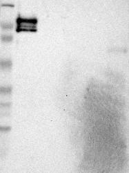
- Western blot analysis of antibody specificity using a routine panel composed of IgG/HSA-depleted human plasma and protein lysates from selected human tissues and cell lines.
- Validation comment
- Band of predicted size in kDa (+/-20%) with additional bands present.
- Primary Ab dilution
- 1:500
- Secondary Ab dilution
- 1:7000
- Lane 1
- Marker [kDa]: 230, 110, 82, 49.3, 32.2, 25.5, 17.6
- Lane 2
- RT-4
- Lane 3
- U-251MG sp
- Lane 4
- Human Plasma
- Lane 5
- Liver
- Lane 6
- Tonsil
- Theoretical target weight
- [kDa] 17

Supportive validation
- Submitted by
- per
- Main image
- Experimental details
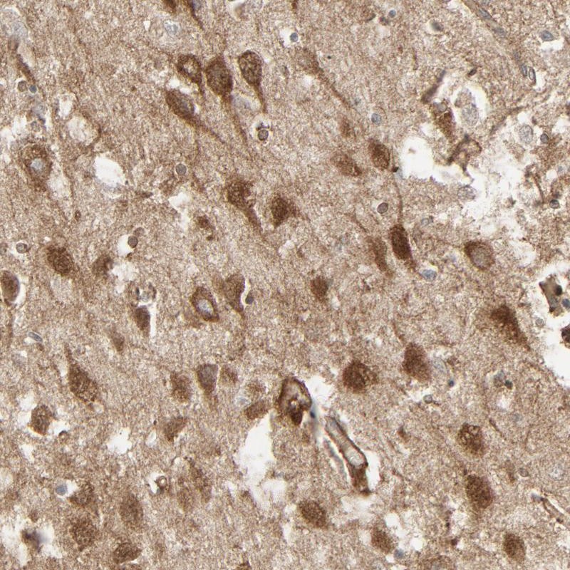
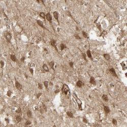
- Immunohistochemical staining of human hippocampus shows strong cytoplasmic positivity in neuronal cells.
- Validation comment
- Staining pattern partly consistent with experimental and/or bioinformatic data.