 Explore
 Validate
 Learn
Learn Western blot
 Immunocytochemistry
Antibody data
- Antibody Data
- Antigen structure
- References [1]
- Comments [0]
- Validations
- Immunocytochemistry [2]
- Immunohistochemistry [1]
Submit
Validation data
Reference
Comment
Report error
- Product number
- MAB7662-100 - Provider product page

- Provider
- R&D Systems
- Product name
- Human FGFR3 (IIIc) Antibody
- Antibody type
- Monoclonal
- Description
- Protein A or G purified from hybridoma culture supernatant. Detects human FGF R3 (IIIc) in direct ELISAs and Western blots. In direct ELISAs, 100% cross-reactivity with with recombinant human (rh) FGF R2 (IIIc), recombinant mouse (rm) FGF R2 (IIIc) and rmFGF R3 (IIIc) is observed. In Western blots (non-reducing conditions only), 50-100% cross-reactivity with rhFGF R2 (IIIc), rmFGF R2 (IIIc) and rmFGF R3 (IIIc) is observed. Does not cross-react with the IIIb isoforms of rhFGF R3, rmFGF R3, rhFGF R2 or rmFGF R2. No cross-reactivity with any isoforms of rhFGF RI or rhFGF R4 is observed.
- Reactivity
- Human
- Host
- Mouse
- Conjugate
- Unconjugated
- Isotype
- IgG
- Antibody clone number
- 136312
- Vial size
- 100 ug
- Storage
- Use a manual defrost freezer and avoid repeated freeze-thaw cycles. 12 months from date of receipt, -20 to -70 °C as supplied. 1 month, 2 to 8 °C under sterile conditions after reconstitution. 6 months, -20 to -70 °C under sterile conditions after reconstitution.
Submitted references Alternative splicing of fibroblast growth factor receptor 3 produces a secreted isoform that inhibits fibroblast growth factor-induced proliferation and is repressed in urothelial carcinoma cell lines.
Tomlinson DC, L'Hôte CG, Kennedy W, Pitt E, Knowles MA
Cancer research 2005 Nov 15;65(22):10441-9
Cancer research 2005 Nov 15;65(22):10441-9
No comments: Submit comment
Supportive validation
- Submitted by
- R&D Systems (provider)
- Main image
- Experimental details
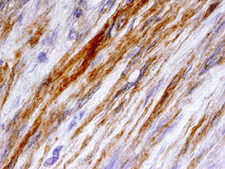
- FGF R3 in Human Bladder Cancer Tissue. FGF R3 was detected in immersion fixed paraffin-embedded sections of human bladder cancer tissue using Mouse Anti-Human FGF R3 (IIIc) Monoclonal Antibody (Catalog # MAB7662) at 25 µg/mL overnight at 4 °C. Tissue was stained using the Anti-Mouse HRP-DAB Cell & Tissue Staining Kit (brown; Catalog # CTS002) and counterstained with hematoxylin (blue). Specific staining was localized to smooth muscle cells. View our protocol for Chromogenic IHC Staining of Paraffin-embedded Tissue Sections.

- Submitted by
- R&D Systems (provider)
- Main image
- Experimental details
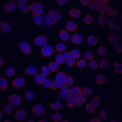
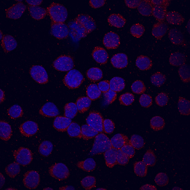
- FGF R3 in U937 Human Cell Line. FGF R3 was detected in immersion fixed U937 human histiocytic lymphoma cell line using Mouse Anti-Human FGF R3 (IIIc) Monoclonal Antibody (Catalog # MAB7662) at 10 µg/mL for 3 hours at room temperature. Cells were stained using the NorthernLights™ 557-conjugated Anti-Mouse IgG Secondary Antibody (red; Catalog # NL007) and counterstained with DAPI (blue). Specific staining was localized to cell surfaces and cytoplasm. View our protocol for Fluorescent ICC Staining of Non-adherent Cells.
Supportive validation
- Submitted by
- R&D Systems (provider)
- Main image
- Experimental details
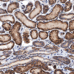
- FGF R3 in Human Kidney. FGF R3 was detected in immersion fixed paraffin-embedded sections of human kidney using Mouse Anti-Human FGF R3 (IIIc) Monoclonal Antibody (Catalog # MAB7662) at 8 µg/mL for 1 hour at room temperature followed by incubation with the Anti-Mouse IgG VisUCyte™ HRP Polymer Antibody (Catalog # VC001). Before incubation with the primary antibody, tissue was subjected to heat-induced epitope retrieval using Antigen Retrieval Reagent-Basic (Catalog # CTS013). Tissue was stained using DAB (brown) and counterstained with hematoxylin (blue). Specific staining was localized to cytoplasm and nuclei. View our protocol for IHC Staining with VisUCyte HRP Polymer Detection Reagents.
