Explore
 Validate
 Learn
Learn Western blot
Antibody data
- Antibody Data
- Antigen structure
- References [0]
- Comments [0]
- Validations
- Western blot [3]
- Immunohistochemistry [2]
Submit
Validation data
Reference
Comment
Report error
- Product number
- TA801078 - Provider product page

- Provider
- Invitrogen Antibodies
- Product name
- FGFR3 Monoclonal Antibody (OTI1B10), TrueMAB™
- Antibody type
- Monoclonal
- Antigen
- Recombinant protein fragment
- Reactivity
- Human
- Host
- Mouse
- Isotype
- IgG
- Antibody clone number
- OTI1B10
- Vial size
- 100 µL
- Concentration
- 1 mg/mL
- Storage
- -20° C, Avoid Freeze/Thaw Cycles
No comments: Submit comment
Supportive validation
- Submitted by
- Invitrogen Antibodies (provider)
- Main image

- Experimental details
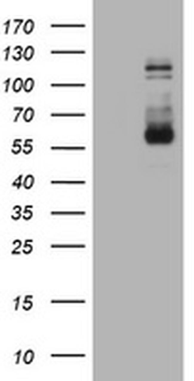
- HEK293T cells were transfected with the pCMV6-ENTRY control (Left lane) or pCMV6-ENTRY FGFR3 (RC215533, Right lane) cDNA for 48 hrs and lysed. Equivalent amounts of cell lysates (5 µg per lane) were separated by SDS-PAGE and immunoblotted with anti-FGFR3. Positive lysates LY424902 (100 µg) and LC424902 (20 µg) can be purchased separately from OriGene.

- Submitted by
- Invitrogen Antibodies (provider)
- Main image

- Experimental details
- HEK293T cells were transfected with the pCMV6-ENTRY control (Left lane) or pCMV6-ENTRY FGFR3 (RC215533, Right lane) cDNA for 48 hrs and lysed. Equivalent amounts of cell lysates (5 µg per lane) were separated by SDS-PAGE and immunoblotted with anti-FGFR3. Positive lysates LY424902 (100 µg) and LC424902 (20 µg) can be purchased separately from OriGene.
- Submitted by
- Invitrogen Antibodies (provider)
- Main image
- Experimental details
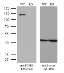
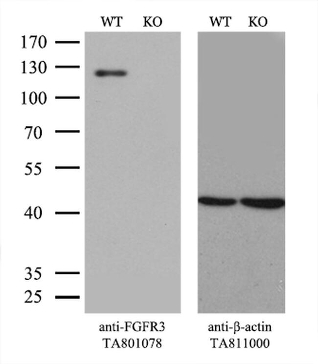
- Equivalent amounts of cell lysates (10 µg per lane) of wild-type 293T cells (WT, Cat# LC810293T) and FGFR3-Knockout 293T cells (KO, Cat# LC810079) were separated by SDS-PAGE and immunoblotted with anti-FGFR3 monoclonal antibody TA801078. Then the blotted membrane was stripped and reprobed with anti-beta-actin antibody (TA811000) as a loading control. (1:500)
Supportive validation
- Submitted by
- Invitrogen Antibodies (provider)
- Main image
- Experimental details
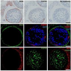
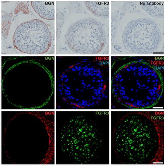
- Figure from citation: Immunohistochemical and immunofluorescence staining with anti-BGN and anti-FGFR3 of serial sections of adult human testis tissue with full spermatogenesis and fetal bone sample (gestational week 10). Scale bar is 50 um for all images. Dilution: 1:200 View Citation
- Submitted by
- Invitrogen Antibodies (provider)
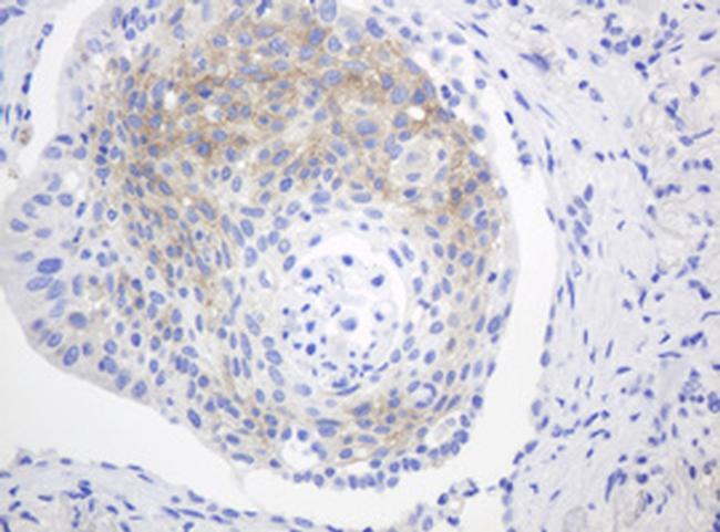
- Main image
- Experimental details
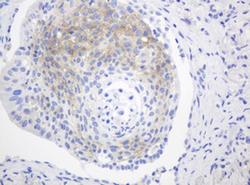
- Immunohistochemical staining of paraffin-embedded Carcinoma of Human lung tissue using anti-FGFR3 mouse monoclonal antibody. (Heat-induced epitope retrieval by 10mM citric buffer, pH6.0, 120°C for 3min, TA801078)