Explore
 Validate
 Learn
Learn Western blot
Antibody data
- Antibody Data
- Antigen structure
- References [6]
- Comments [0]
- Validations
- Western blot [4]
- Immunocytochemistry [1]
- Immunoprecipitation [1]
- Immunohistochemistry [2]
Submit
Validation data
Reference
Comment
Report error
- Product number
- GTX101820 - Provider product page

- Provider
- GeneTex
- Proper citation
- GeneTex Cat#GTX101820, RRID:AB_1241460
- Product name
- Ku70 antibody
- Antibody type
- Polyclonal
- Reactivity
- Human
- Host
- Rabbit
Submitted references Chloroquine inhibits human retina pigmented epithelial cell growth and microtubule nucleation by downregulating p150glued.
7-hydroxy-staurosporine, UCN-01, induces DNA damage response, and autophagy in human osteosarcoma U2-OS cells.
CD24+ tumor-initiating cells from oral squamous cell carcinoma induce initial angiogenesis in vivo.
Overexpression of TNKS1BP1 in lung cancers and its involvement in homologous recombination pathway of DNA double-strand breaks.
Classical non-homologous end-joining pathway utilizes nascent RNA for error-free double-strand break repair of transcribed genes.
NR5A1 prevents centriole splitting by inhibiting centrosomal DNA-PK activation and β-catenin accumulation.
Chen TY, Lien WC, Cheng HL, Kuan TS, Sheu SY, Wang CY
Journal of cellular physiology 2019 Jul;234(7):10445-10457
Journal of cellular physiology 2019 Jul;234(7):10445-10457
7-hydroxy-staurosporine, UCN-01, induces DNA damage response, and autophagy in human osteosarcoma U2-OS cells.
Lien WC, Chen TY, Sheu SY, Lin TC, Kang FC, Yu CH, Kuan TS, Huang BM, Wang CY
Journal of cellular biochemistry 2018 Jun;119(6):4729-4741
Journal of cellular biochemistry 2018 Jun;119(6):4729-4741
CD24+ tumor-initiating cells from oral squamous cell carcinoma induce initial angiogenesis in vivo.
Zimmerer RM, Ludwig N, Kampmann A, Bittermann G, Spalthoff S, Jungheim M, Gellrich NC, Tavassol F
Microvascular research 2017 Jul;112:101-108
Microvascular research 2017 Jul;112:101-108
Overexpression of TNKS1BP1 in lung cancers and its involvement in homologous recombination pathway of DNA double-strand breaks.
Tan W, Guan H, Zou LH, Wang Y, Liu XD, Rang WQ, Zhou PK, Pei HD, Zhong CG
Cancer medicine 2017 Feb;6(2):483-493
Cancer medicine 2017 Feb;6(2):483-493
Classical non-homologous end-joining pathway utilizes nascent RNA for error-free double-strand break repair of transcribed genes.
Chakraborty A, Tapryal N, Venkova T, Horikoshi N, Pandita RK, Sarker AH, Sarkar PS, Pandita TK, Hazra TK
Nature communications 2016 Oct 5;7:13049
Nature communications 2016 Oct 5;7:13049
NR5A1 prevents centriole splitting by inhibiting centrosomal DNA-PK activation and β-catenin accumulation.
Wang CY, Lai PY, Chen TY, Chung BC
Cell communication and signaling : CCS 2014 Nov 25;12:55
Cell communication and signaling : CCS 2014 Nov 25;12:55
No comments: Submit comment
Supportive validation
- Submitted by
- GeneTex (provider)
- Main image
- Experimental details
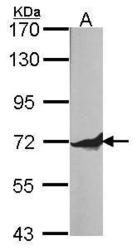
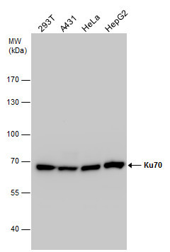
- Sample (30 ug of whole cell lysate) A: A431 (GTX27909) 7.5% SDS PAGE Ku-70 antibody GTX101820 diluted at 1:500
- Validation comment
- WB

- Submitted by
- GeneTex (provider)
- Main image
- Experimental details
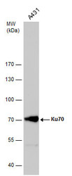
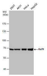
- Ku70 antibody detects Ku70 protein by western blot analysis. Whole cell extracts (30 ?g) was separated by 7.5% SDS-PAGE, and the membrane was blotted with Ku70 antibody (GTX101820) diluted by 1:1000.
- Validation comment
- WB
- Submitted by
- GeneTex (provider)
- Main image
- Experimental details
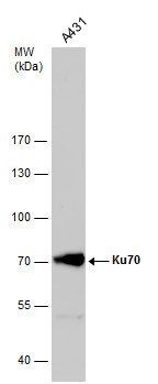
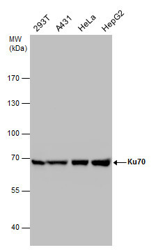
- Ku70 antibody detects Ku70 protein by western blot analysis. Various whole cell extracts (30 ?g) were separated by 7.5% SDS-PAGE, and the membrane was blotted with Ku70 antibody (GTX101820) diluted at a dilution of 1:2000.
- Validation comment
- WB
- Submitted by
- GeneTex (provider)
- Main image
- Experimental details
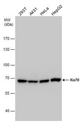
- Ku70 antibody detects Ku70 protein by western blot analysis. Various whole cell extracts (30 ?g) were separated by 7.5% SDS-PAGE, and the membrane was blotted with Ku70 antibody (GTX101820) diluted by 1:2000. The HRP-conjugated anti-rabbit IgG antibody (GTX213110-01) was used to detect the primary antibody.
Supportive validation
- Submitted by
- GeneTex (provider)
- Main image
- Experimental details
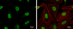
- Ku70 antibody detects Ku70 protein at nucleus by immunofluorescent analysis. Samples: HeLa cells were fixed in 4% paraformaldehyde at RT for 15 min.Green: Ku70 protein stained by Ku70 antibody (GTX101820) diluted at 1:200.Red: phalloidin, a cytoskeleton marker, diluted at 1:200.Scale bar = 10 £gm.

Supportive validation
- Submitted by
- GeneTex (provider)
- Main image
- Experimental details
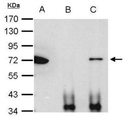
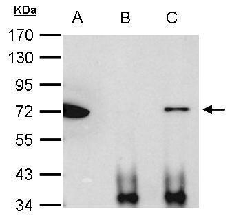
- Ku70 antibody immunoprecipitates Ku70 protein in IP experiments. IP Sample: 1000 ?g HeLa whole cell lysate/extract A. 40 £gg HeLa whole cell lysate/extract B. Control with 2.5 £gg of preimmune rabbit IgG C. Immunoprecipitation of Ku70 protein by 2.5 £gg of Ku70 antibody (GTX101820) 7.5% SDS-PAGE The immunoprecipitated Ku70 protein was detected by Ku70 antibody (GTX101820) diluted at 1:1000. EasyBlot anti-rabbit IgG (GTX221666-01) was used as a secondary reagent.
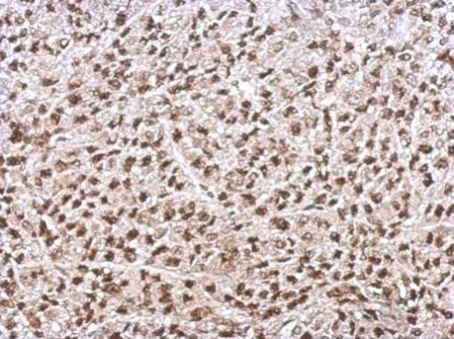
Supportive validation
- Submitted by
- GeneTex (provider)
- Main image
- Experimental details
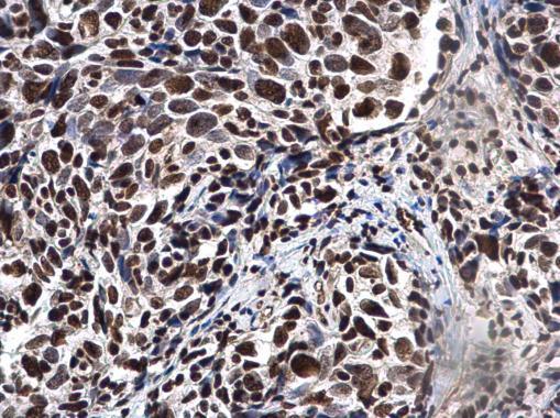
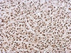
- Immunohistochemical analysis of paraffin-embedded D54 xenograft, using Ku70(GTX101820) antibody at 1:500 dilution.
- Submitted by
- GeneTex (provider)
- Main image
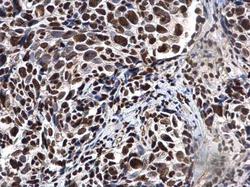
- Experimental details
- Ku70 antibody detects Ku70 protein at nucleus in human cervical cancer by immunohistochemical analysis. Sample: Paraffin-embedded human cervical cancer. Ku70 antibody (GTX101820) diluted at 1:500.