Explore
 Validate
 Learn
Learn Western blot
 ELISA
ELISAAntibody data
- Antibody Data
- Antigen structure
- References [0]
- Comments [0]
- Validations
- Western blot [2]
- Immunocytochemistry [2]
- Immunohistochemistry [1]
- Flow cytometry [1]
Submit
Validation data
Reference
Comment
Report error
- Product number
- F49015 - Provider product page

- Provider
- NSJ Bioreagents
- Product name
- XRCC6 Antibody
- Antibody type
- Polyclonal
- Description
- This highly specific XRCC6 antibody is suitable for use in Western blot/Immunohistochemistry/Flow cytometry/Immunofluorescence/ELISA applications with human samples.
- Reactivity
- Human
- Host
- Rabbit
- Conjugate
- Unconjugated
- Vial size
- 0.08 ml, 0.4 ml
- Concentration
- In 1X PBS, pH 7.4, with 0.09% sodium azide
- Storage
- Aliquot the XRCC6 antibody and store frozen at -20oC or colder. Avoid repeated freeze-thaw cycles.
No comments: Submit comment
Supportive validation
- Submitted by
- NSJ Bioreagents (provider)
- Main image

- Experimental details
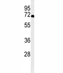
- Western blot analysis of XRCC6 antibody and A2058 lysate.
- Submitted by
- NSJ Bioreagents (provider)
- Main image

- Experimental details
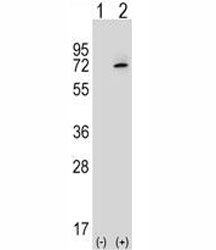
- Western blot analysis of XRCC6 antibody and 293 cell lysate either nontransfected (Lane 1) or transiently transfected (2) with the XRCC6 gene.
Supportive validation
- Submitted by
- NSJ Bioreagents (provider)
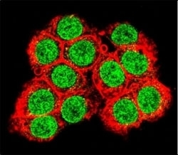
- Main image
- Experimental details
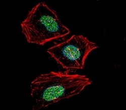
- Fluorescent confocal image of HeLa cells stained with XRCC6 antibody. Alexa Fluor 488 secondary (green) was used. XRCC6 immunoreactivity is localized to the nucleus strongly and cytoplasm weakly.
- Submitted by
- NSJ Bioreagents (provider)
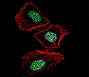
- Main image
- Experimental details
- Confocal immunofluorescent analysis of XRCC6 antibody with 293 cells followed by Alexa Fluor 488-conjugated goat anti-rabbit lgG (green). Actin filaments have been labeled with Alexa Fluor 555 Phalloidin (red).

Supportive validation
- Submitted by
- NSJ Bioreagents (provider)
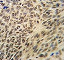
- Main image

- Experimental details
- XRCC6 antibody IHC analysis in formalin fixed and paraffin embedded human lung carcinoma.
Supportive validation
- Submitted by
- NSJ Bioreagents (provider)
- Main image
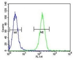
- Experimental details
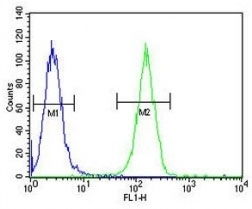
- XRCC6 antibody flow cytometric analysis of A2058 cells (green) compared to a negative control (blue). FITC-conjugated goat-anti-rabbit secondary Ab was used for the analysis.