 Explore
 Validate
 Learn
Learn Western blot
Antibody data
- Antibody Data
- Antigen structure
- References [0]
- Comments [0]
- Validations
- Western blot [5]
- Immunocytochemistry [2]
- Immunohistochemistry [1]
- Flow cytometry [1]
Submit
Validation data
Reference
Comment
Report error
- Product number
- PA5-25011 - Provider product page

- Provider
- Invitrogen Antibodies
- Product name
- Ku70 Polyclonal Antibody
- Antibody type
- Polyclonal
- Antigen
- Synthetic peptide
- Reactivity
- Human
- Host
- Rabbit
- Isotype
- IgG
- Vial size
- 400 µL
- Storage
- Store at 4°C short term. For long term storage, store at -20°C, avoiding freeze/thaw cycles.
No comments: Submit comment
Supportive validation
- Submitted by
- Invitrogen Antibodies (provider)
- Main image

- Experimental details
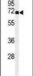
- Western blot analysis of XRCC6 (arrow) using a XRCC6 polyclonal antibody (Product # PA5-25011) in 293 cell lysates (2 µg/lane) either nontransfected (Lane 1) or transiently transfected (Lane 2) with the XRCC6 gene.

- Submitted by
- Invitrogen Antibodies (provider)
- Main image
- Experimental details
- Western blot analysis using a XRCC6 polyclonal antibody (Product # PA5-25011) in A2058 cell lysates (35 µg per lane).

- Submitted by
- Invitrogen Antibodies (provider)
- Main image

- Experimental details
- Western blot analysis of XRCC6 (arrow) using a XRCC6 polyclonal antibody (Product # PA5-25011) in 293 cell lysates (2 µg/lane) either nontransfected (Lane 1) or transiently transfected (Lane 2) with the XRCC6 gene.

- Submitted by
- Invitrogen Antibodies (provider)
- Main image
- Experimental details
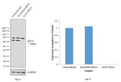
- Knockdown of Ku70 was achieved by transfecting HeLa cells with Ku70 specific siRNAs (Silencer® select Product # s5457, s5455). Western blot analysis (Fig. a) was performed using modified whole cell extracts from the Ku70 knockdown cells (lane 3), non-specific scrambled siRNA transfected cells (lane 2) and untransfected cells (lane 1). The blots were probed with Anti-Ku70 Polyclonal Antibody (Product # PA5-25011, 1:1000 dilution) and Goat anti-Rabbit IgG (H+L) Superclonal™ Secondary Antibody, HRP conjugate (Product # A27036, 0.25 µg/mL, 1:4000 dilution). Densitometric analysis of this western blot is shown in histogram (Fig. b). Decrease in signal upon siRNA mediated knock down confirms that antibody is specific to Ku70. An additional band was also observed at ~80kDa.

- Submitted by
- Invitrogen Antibodies (provider)
- Main image

- Experimental details
- Western blot analysis was performed on modified whole cell extracts (1% SDS) (30 µg lysate) of HeLa (Lane 1), K562 (Lane 2), Jurkat (Lane 3), HEK293 (Lane 4) and HepG2 (Lane 5). The blot was probed with Anti-Ku70 Polyclonal Antibody (Product # PA5-25011, 1:1000 dilution) and detected by chemiluminescence using Goat anti-Rabbit IgG (H+L) Superclonal™ Secondary Antibody, HRP conjugate (Product # A27036, 0.25 µg/mL, 1:4000 dilution). A 70kDa band corresponding to Ku70 was seen in all cell lines tested. An additional band was also observed at ~80kDa across the cell lines.

Supportive validation
- Submitted by
- Invitrogen Antibodies (provider)
- Main image
- Experimental details
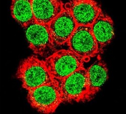
- Immunofluorescent analysis of 293 cells using a XRCC6 polyclonal antibody (Product # PA5-25011) at a dilution of 1:10-50. Primary antibody was detected with goat anti-rabbit lgG, fluor-conjugated secondary antibody (green). Actin filaments have been labeled with red dye conjugated phalloidin.

- Submitted by
- Invitrogen Antibodies (provider)
- Main image
- Experimental details
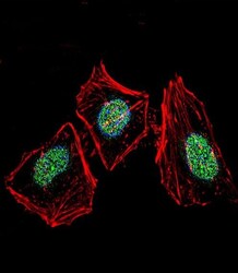
- Immunofluorescent analysis of HeLa cells using a XRCC6 polyclonal antibody (Product # PA5-25011). HeLa cells were fixed with 4% PFA (20 min), permeabilized with Triton X-100 (0.1%, 10 min), then incubated with a XRCC6 polyclonal antibody (Product # PA5-25011) (1:25, 1 hr at 37°C). Primary antibody was detected with fluor-conjugated donkey anti-rabbit secondary antibody (green) at 1:400 dilution for 50 min at 37°C). Actin filaments have been labeled with dye-conjugated phalloidin (red). Nuclei were counterstained with DAPI (blue) (10 µg/mL, 10 min).

Supportive validation
- Submitted by
- Invitrogen Antibodies (provider)
- Main image
- Experimental details
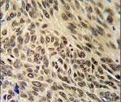
- Immunohistochemistry analysis in formalin-fixed, paraffin-embedded human lung carcinoma using a XRCC6 polyclonal antibody (Product # PA5-25011), followed by HRP-conjugated secondary antibody and DAB staining.

Supportive validation
- Submitted by
- Invitrogen Antibodies (provider)
- Main image
- Experimental details
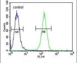
- Flow cytometry analysis of A2058 cells using a XRCC6 polyclonal antibody (Product # PA5-25011) (right) compared to a negative control cell (left) at a dilution of 1:10-50, followed by a FITC-conjugated goat anti-rabbit antibody
