Explore
 Validate
 Learn
LearnMA5-14877
antibody from Invitrogen Antibodies
Targeting: SUMO1
GMP1, OFC10, PIC1, SMT3C, SMT3H3, SUMO-1, UBL1
 Western blot
 Immunocytochemistry
Antibody data
- Antibody Data
- Antigen structure
- References [0]
- Comments [0]
- Validations
- Immunocytochemistry [5]
- Immunohistochemistry [1]
Submit
Validation data
Reference
Comment
Report error
- Product number
- MA5-14877 - Provider product page

- Provider
- Invitrogen Antibodies
- Product name
- SUMO1 Monoclonal Antibody (T.243.0)
- Antibody type
- Monoclonal
- Antigen
- Synthetic peptide
- Description
- It is not recommended to aliquot this antibody.
- Reactivity
- Human, Mouse, Rat
- Host
- Rabbit
- Isotype
- IgG
- Antibody clone number
- T.243.0
- Vial size
- 100 µL
- Concentration
- 23 µg/mL
- Storage
- -20°C
No comments: Submit comment
Supportive validation
- Submitted by
- Invitrogen Antibodies (provider)
- Main image
- Experimental details
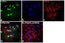
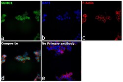
- Immunofluorescence analysis of SUMO1 (Small ubiquitin-related modifier 1) was performed using 80% confluent log phase Hep G2 cells. The cells were fixed with 4% paraformaldehyde for 10 minutes, permeabilized with 0.1% Triton™ X-100 for 10 minutes, and blocked with 2% BSA for 1 hour at room temperature. The cells were labeled with SUMO1 Monoclonal Antibody (T.243.0) (Product # MA5-14877) at 1:100 in 0.1% BSA, incubated at 4 degree celsius overnight and then labeled with Goat anti-Rabbit IgG (H+L) Highly Cross-Adsorbed Secondary Antibody, Alexa Fluor Plus 488 (Product # A32731), (1:2000), for 45 minutes at room temperature (Panel a: Green). Nuclei (Panel b:Blue) were stained with Hoechst 33342 (Product # H1399). F-actin (Panel c: Red) was stained with Rhodamine Phalloidin (Product # R415, 1:300). Panel d represents the merged image showing punctate structures within the nucleus and nucleoplasm localization for SUMO1. Panel e represents control cells with no primary antibody to assess background. The images were captured at 40X magnification with CellInsight CX7 LZR High-Content Screening (HCS) Platform (Product # CX7C1115LZR)
- Submitted by
- Invitrogen Antibodies (provider)
- Main image
- Experimental details
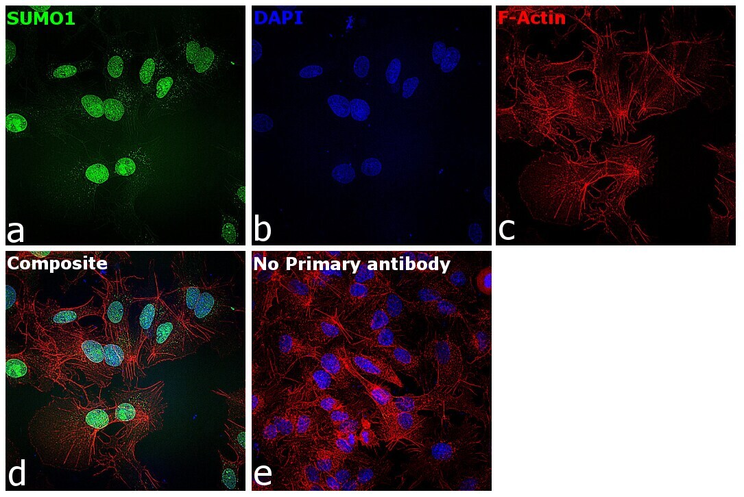
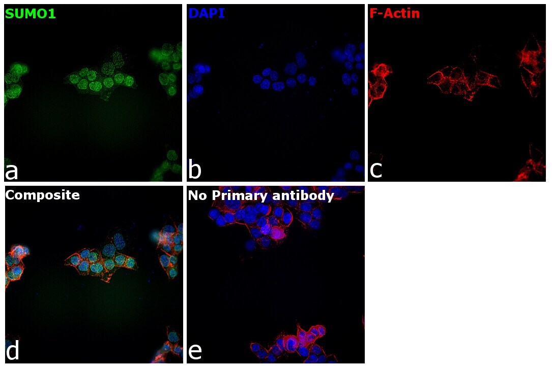
- Immunofluorescence analysis of SUMO1 (Small ubiquitin-related modifier 1) was performed using 80% confluent log phase A-431 cells. The cells were fixed with 4% paraformaldehyde for 10 minutes, permeabilized with 0.1% Triton™ X-100 for 10 minutes, and blocked with 2% BSA for 1 hour at room temperature. The cells were labeled with SUMO1 Monoclonal Antibody (T.243.0) (Product # MA5-14877) at 1:100 in 0.1% BSA, incubated at 4 degree celsius overnight and then labeled with Goat anti-Rabbit IgG (H+L) Highly Cross-Adsorbed Secondary Antibody, Alexa Fluor Plus 488 (Product # A32731), (1:2,000), for 45 minutes at room temperature (Panel a: Green). Nuclei (Panel b:Blue) were stained with Hoechst 33342 (Product # H1399). F-actin (Panel c: Red) was stained with Rhodamine Phalloidin (Product # R415, 1:300). Panel d represents the merged image showing punctate structures within the nucleus and nucleoplasm localization for SUMO1. Panel e represents control cells with no primary antibody to assess background. The images were captured at 40X magnification with CellInsight CX7 LZR High-Content Screening (HCS) Platform (Product # CX7C1115LZR).

- Submitted by
- Invitrogen Antibodies (provider)
- Main image
- Experimental details
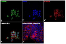
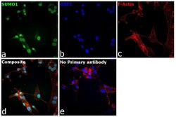
- Immunofluorescence analysis of SUMO1 (Small ubiquitin-related modifier 1) was performed using 80% confluent log phase U-2 OS cells. The cells were fixed with 4% paraformaldehyde for 10 minutes, permeabilized with 0.1% Triton™ X-100 for 10 minutes, and blocked with 2% BSA for 1 hour at room temperature. The cells were labeled with SUMO1 Monoclonal Antibody (T.243.0) (Product # MA5-14877) at 1:100 in 0.1% BSA, incubated at 4 degree celsius overnight and then labeled with Goat anti-Rabbit IgG (H+L) Highly Cross-Adsorbed Secondary Antibody, Alexa Fluor Plus 488 (Product # A32731), (1:2,000), for 45 minutes at room temperature (Panel a: Green). Nuclei (Panel b:Blue) were stained with Hoechst 33342 (Product # H1399). F-actin (Panel c: Red) was stained with Rhodamine Phalloidin (Product # R415, 1:300). Panel d represents the merged image showing punctate structures within the nucleus and nucleoplasm localization for SUMO1. Panel e represents control cells with no primary antibody to assess background. The images were captured at 40X magnification with CellInsight CX7 LZR High-Content Screening (HCS) Platform (Product # CX7C1115LZR).
- Submitted by
- Invitrogen Antibodies (provider)
- Main image
- Experimental details
- Immunofluorescence analysis of SUMO1 (Small ubiquitin-related modifier 1) was performed using 80% confluent log phase U-87 MG cells. The cells were fixed with 4% paraformaldehyde for 10 minutes, permeabilized with 0.1% Triton™ X-100 for 10 minutes, and blocked with 2% BSA for 1 hour at room temperature. The cells were labeled with SUMO1 Monoclonal Antibody (T.243.0) (Product # MA5-14877) at 1:100 in 0.1% BSA, incubated at 4 degree celsius overnight and then labeled with Goat anti-Rabbit IgG (H+L) Highly Cross-Adsorbed Secondary Antibody, Alexa Fluor Plus 488 (Product # A32731), (1:2,000), for 45 minutes at room temperature (Panel a: Green). Nuclei (Panel b:Blue) were stained with Hoechst 33342 (Product # H1399). F-actin (Panel c: Red) was stained with Rhodamine Phalloidin (Product # R415, 1:300). Panel d represents the merged image showing punctate structures within the nucleus and nucleoplasm localization for SUMO1. Panel e represents control cells with no primary antibody to assess background. The images were captured at 40X magnification with CellInsight CX7 LZR High-Content Screening (HCS) Platform (Product # CX7C1115LZR).

- Submitted by
- Invitrogen Antibodies (provider)
- Main image

- Experimental details
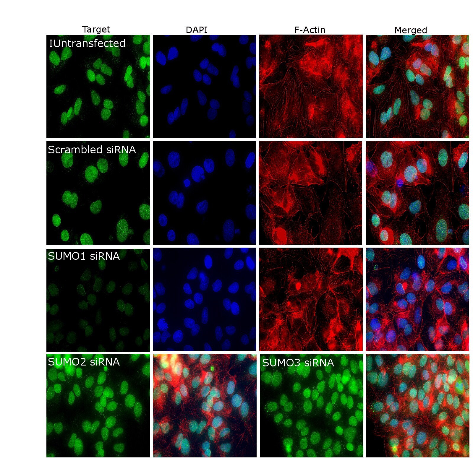
- Knockdown of SUMO1 was achieved by transfecting Hep G2 cells with SUMO1 specific siRNA (Silencer® select Product # s14608 , s14609). Immunofluorescence analysis was performed on untransfected Hep G2 cells, transfected with non-specific scrambled siRNA, transfected with SUMO1 specific siRNA, SUMO2 specific siRNA and SUMO3 specific siRNA. Cells were fixed, permeabilized, and labelled with SUMO1 Monoclonal Antibody (T.243.0) (Product # MA5-14877, 1:100) followed by Donkey anti-Rabbit IgG (H+L) Highly Cross-Adsorbed Secondary Antibody, Alexa Fluor Plus 488 (Product # A32790), (1:2500 dilution). Nuclei (blue) were stained using ProLong™ Diamond Antifade Mountant with DAPI (Product # P36962), and Rhodamine Phalloidin (Product # R415, 1:300) was used for cytoskeletal F-actin (Red) staining. Reduction of specific signal was observed only in SUMO1 siRNA transfected cells but not in SUMO2 or SUMO3 transfected cells, confirming specificity of the antibody to SUMO1 (Green). The Images were captured at 60X magnification with EVOS™ M7000 Imaging System (Product # AMF7000).
Supportive validation
- Submitted by
- Invitrogen Antibodies (provider)
- Main image
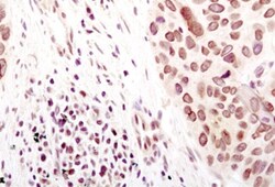
- Experimental details
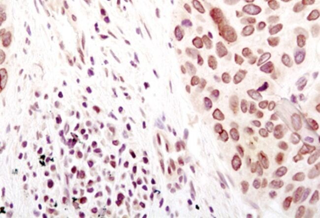
- Immunohistochemical analysis of SUMO-1 in paraffin-embedded human lung carcinoma using a SUMO-1 monoclonal antibody (Product # MA5-14877) in the presence of control peptide (left) or antigen specific peptide (right).