Explore
 Validate
 Learn
Learn Western blot
 Immunocytochemistry
Antibody data
- Antibody Data
- Antigen structure
- References [2]
- Comments [0]
- Validations
- Immunocytochemistry [2]
- Flow cytometry [1]
Submit
Validation data
Reference
Comment
Report error
- Product number
- 700044 - Provider product page

- Provider
- Invitrogen Antibodies
- Product name
- Phospho-PLCG1 (Tyr783) Recombinant Rabbit Monoclonal Antibody (C-p1p2-39H16L83)
- Antibody type
- Monoclonal
- Antigen
- Synthetic peptide
- Description
- This antibody is predicted to react with bovine, mouse, rat, Xenopus and zebrafish based on sequence homology. Intact IgG appears on a non-reducing gel as ~150 kDa band and upon reduction generating a ~25 kDa light chain band and a ~50 kDa heavy chain. Recombinant rabbit monoclonal antibodies are produced using in vitro expression systems. The expression systems are developed by cloning in the specific antibody DNA sequences from immunoreactive rabbits. Then, individual clones are screened to select the best candidates for production. The advantages of using recombinant rabbit monoclonal antibodies include: better specificity and sensitivity, lot-to-lot consistency, animal origin-free formulations, and broader immunoreactivity to diverse targets due to larger rabbit immune repertoire.
- Reactivity
- Human, Mouse
- Host
- Rabbit
- Isotype
- IgG
- Antibody clone number
- C-p1p2-39H16L83
- Vial size
- 100 μg
- Concentration
- 0.5 mg/mL
- Storage
- Store at 4°C short term. For long term storage, store at -20°C, avoiding freeze/thaw cycles.
Submitted references Quantitative differences in CD45 expression unmask functions for CD45 in B-cell development, tolerance, and survival.
Glucocorticoid receptors recruit the CaMKIIα-BDNF-CREB pathways to mediate memory consolidation.
Zikherman J, Doan K, Parameswaran R, Raschke W, Weiss A
Proceedings of the National Academy of Sciences of the United States of America 2012 Jan 3;109(1):E3-12
Proceedings of the National Academy of Sciences of the United States of America 2012 Jan 3;109(1):E3-12
Glucocorticoid receptors recruit the CaMKIIα-BDNF-CREB pathways to mediate memory consolidation.
Chen DY, Bambah-Mukku D, Pollonini G, Alberini CM
Nature neuroscience 2012 Dec;15(12):1707-14
Nature neuroscience 2012 Dec;15(12):1707-14
No comments: Submit comment
Supportive validation
- Submitted by
- Invitrogen Antibodies (provider)
- Main image

- Experimental details
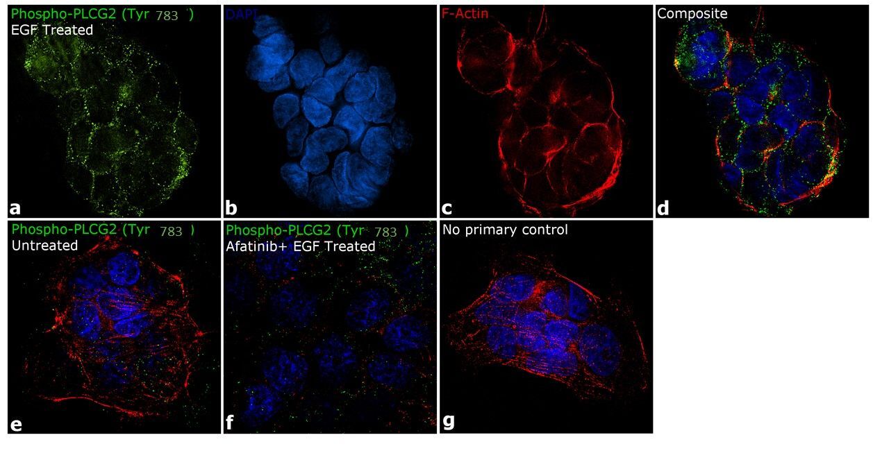
- Immunofluorescence analysis of Phospho-PLCG1 (Tyr783) was performed using 70% confluent log phase A-431 cells treated with 200 ng/mL of EGF for 10 minutes. The cells were fixed with 4% paraformaldehyde for 10 minutes, permeabilized with 0.1% Triton™ X-100 for 10 minutes and blocked with 1% BSA for 1 hour at room temperature. The cells were labeled with Phospho-PLCG1 (Tyr783) Antibody (C-p1p2-39H16L83), Recombinant Rabbit Monoclonal (Product # 700044) at 2 µg/mL in 0.1% BSA and incubated overnight at 4°C and then labeled with Goat anti-Rabbit IgG (H+L) Superclonal™ Secondary Antibody, Alexa Fluor® 488 conjugate (Product # A27034) at a dilution of 1:2,000 for 45 minutes at room temperature (Panel a: green). Nuclei (Panel b: blue) were stained with SlowFade® Gold Antifade Mountant with DAPI (Product # S36938). F-actin (Panel c: red) was stained with Rhodamine Phalloidin (Product # R415, 1:300). Panel d represents the merged image showing membrane localization. Panel f represents cells treated with antagonist, Afatinib (1 µM for 6 hrs) followed by EGF (200 ng/mL for 10 minutes), showing reduced Phospho-PLCG2 (Tyr1217) staining. Panel e shows untreated cells with no signal. Panel g represents control cells with no primary antibody to assess background. The images were captured at 60X magnification.
- Submitted by
- Invitrogen Antibodies (provider)
- Main image

- Experimental details
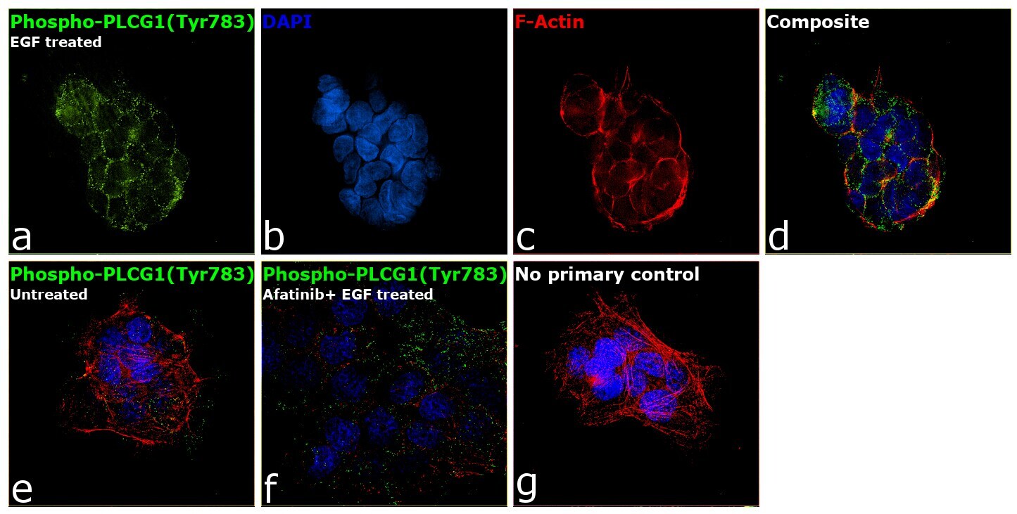
- Immunofluorescence analysis of Phospho-PLCG1 (Tyr783) was performed using 70% confluent log phase A-431 cells treated with 200 ng/mL of EGF for 10 minutes. The cells were fixed with 4% paraformaldehyde for 10 minutes, permeabilized with 0.1% Triton™ X-100 for 10 minutes and blocked with 1% BSA for 1 hour at room temperature. The cells were labeled with Phospho-PLCG1 (Tyr783) Antibody (C-p1p2-39H16L83), Recombinant Rabbit Monoclonal (Product # 700044) at 2 µg/mL in 0.1% BSA and incubated overnight at 4°C and then labeled with Goat anti-Rabbit IgG (Heavy Chain) Superclonal™ Secondary Antibody, Alexa Fluor® 488 conjugate (Product # A27034) at a dilution of 1:2,000 for 45 minutes at room temperature (Panel a: green). Nuclei (Panel b: blue) were stained with SlowFade® Gold Antifade Mountant with DAPI (Product # S36938). F-actin (Panel c: red) was stained with Rhodamine Phalloidin (Product # R415, 1:300). Panel d represents the merged image showing membrane localization. Panel f represents cells treated with antagonist, Afatinib (1 µM for 6 hrs) followed by EGF (200 ng/mL for 10 minutes), showing reduced Phospho-PLCG2 (Tyr1217) staining. Panel e shows untreated cells with no signal. Panel g represents control cells with no primary antibody to assess background. The images were captured at 60X magnification.
Supportive validation
- Submitted by
- Invitrogen Antibodies (provider)
- Main image
- Experimental details
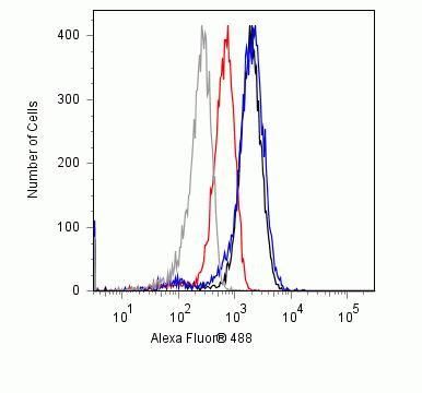
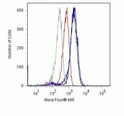
- Flow cytometry analysis of Phospho-PLC gamma pTyr783 in NIH-3T3 cells using a Phospho-PLC gamma pTyr783 recombinant rabbit monoclonal antibody (Product # 700044) at a dilution of 3ug. Cells were fixed and permeabilized using FIX & PERM (Product # GAS004) reagent, and detection was performed using an Alexa Fluor 488 goat anti-rabbit IgG (black) compared to a control without primary antibody (gray). Samples were preincubated for 30 minutes in the presence of 50ug/ml of the specific phosphopeptide (red) as well as the non-phosphopeptide (blue) to show specificity for the phosphopeptide only.