 Explore
 Validate
 Learn
Learn419300
antibody from Invitrogen Antibodies
Targeting: PPARG
NR1C3, PPARG1, PPARG2, PPARgamma
 Western blot
 ELISA Immunoprecipitation Immunohistochemistry Gel shift Chromatin Immunoprecipitation Other assay
Antibody data
- Antibody Data
- Antigen structure
- References [1]
- Comments [0]
- Validations
- Western blot [3]
- Immunohistochemistry [2]
- Other assay [1]
Submit
Validation data
Reference
Comment
Report error
- Product number
- 419300 - Provider product page

- Provider
- Invitrogen Antibodies
- Product name
- PPAR gamma Monoclonal Antibody (A3409A)
- Antibody type
- Monoclonal
- Antigen
- Recombinant full-length protein
- Description
- This antibody specifically recognizes human PPAR gamma1 and 2 and cross reacts with mouse and rat PPAR gamma1 and 2. This antibody does not recognize human PPAR alpha and delta.
- Reactivity
- Human, Mouse, Rat
- Host
- Mouse
- Isotype
- IgG
- Antibody clone number
- A3409A
- Vial size
- 100 µL
- Concentration
- 1 mg/mL
- Storage
- Maintain refrigerated at 2-8°C for up to 1 month. For long term storage store at -20°C
Submitted references Unacylated ghrelin promotes adipogenesis in rodent bone marrow via ghrelin O-acyl transferase and GHS-R(1a) activity: evidence for target cell-induced acylation.
Hopkins AL, Nelson TA, Guschina IA, Parsons LC, Lewis CL, Brown RC, Christian HC, Davies JS, Wells T
Scientific reports 2017 Mar 31;7:45541
Scientific reports 2017 Mar 31;7:45541
No comments: Submit comment
Supportive validation
- Submitted by
- Invitrogen Antibodies (provider)
- Main image

- Experimental details
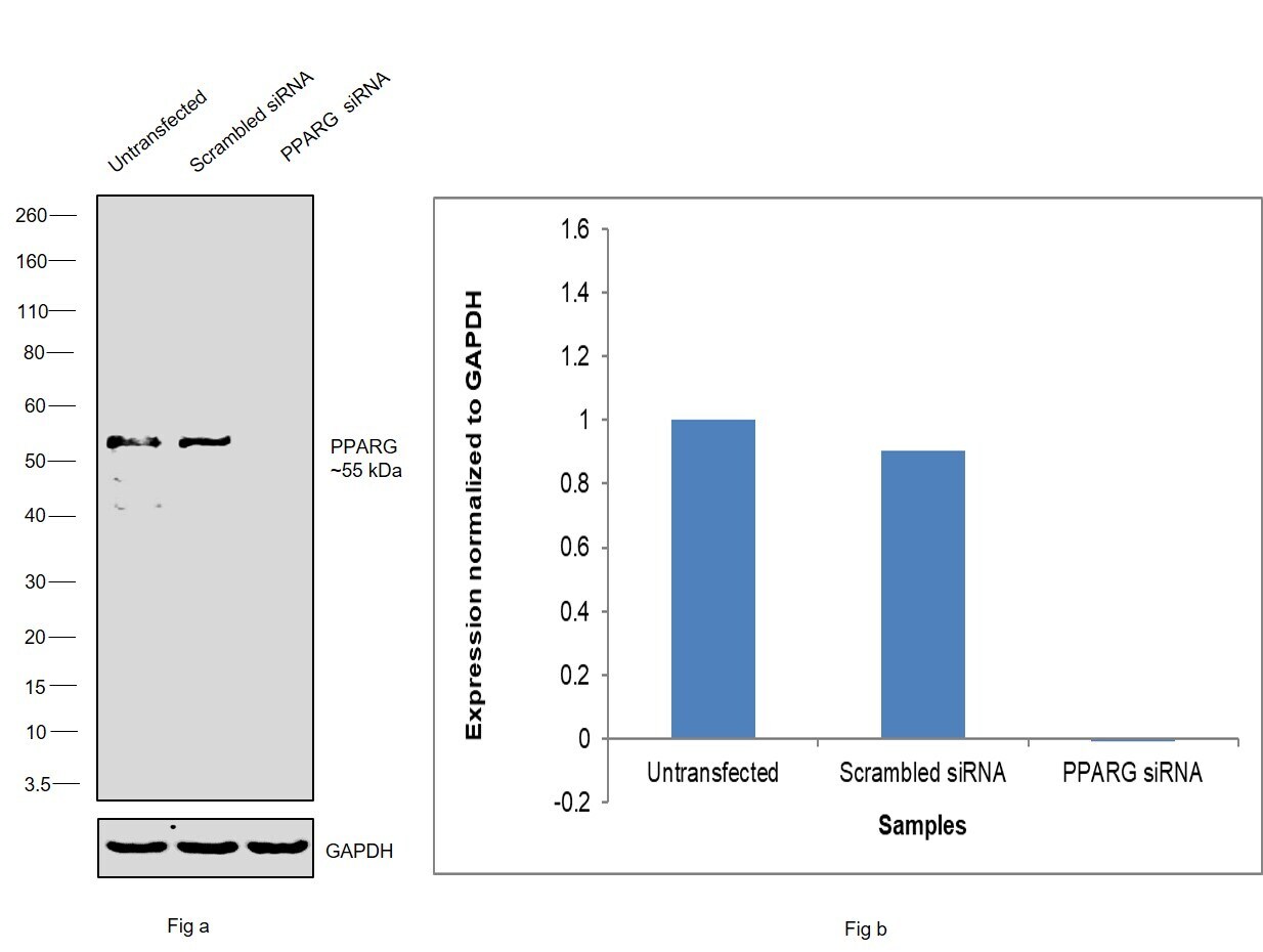
- Knockdown of Peroxisome proliferator-activated receptor gamma was achieved by transfecting A549 with Peroxisome proliferator-activated receptor gamma specific siRNAs (Silencer® select Product # S10888, S10887). Western blot analysis (Fig. a) was performed using Whole cell extracts from the Peroxisome proliferator-activated receptor gamma knockdown cells (lane 3), non-targeting scrambled siRNA transfected cells (lane 2) and untransfected cells (lane 1). The blot was probed with PPAR gamma Monoclonal Antibody (A3409A) (Product # 419300, 1 µg/mL ) and Goat anti-Mouse IgG (H+L) Superclonal™ Recombinant Secondary Antibody, HRP (Product # A28177, 1:4000 dilution). Densitometric analysis of this western blot is shown in histogram (Fig. b). Decrease in signal upon siRNA mediated knock down confirms that antibody is specific to Peroxisome proliferator-activated receptor gamma.
- Submitted by
- Invitrogen Antibodies (provider)
- Main image
- Experimental details
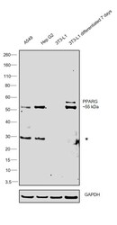
- Western blot was performed using Anti-PPAR gamma Monoclonal Antibody (A3409A) (Product # 419300) and two bands at ~55kDa and ~ 57 kDa corresponding to Peroxisome proliferator-activated receptor gamma was observed across cell lines tested. Whole cell extracts (30 µg lysate) of A549 (Lane 1), Hep G2 (Lane 2), 3T3-L1 (Lane 3), 3T3-L1 differentiated for 7 days (Lane 4) were electrophoresed using NuPAGE™ 4-12% Bis-Tris Protein Gel (Product # NP0321BOX). Resolved proteins were then transferred onto a Nitrocellulose membrane (Product # LC2001) by iBlot® 2 Dry Blotting System (Product # IB21001). The blot was probed with the primary antibody (1 µg/mL) and detected by chemiluminescence with Goat anti-Mouse IgG (H+L) Superclonal™ Recombinant Secondary Antibody, HRP (Product # A28177, 1:4000 dilution) using the iBright FL 1000 (Product # A32752). Chemiluminescent detection was performed using Novex® ECL Reagent Kit (Product # WP20005).

- Submitted by
- Invitrogen Antibodies (provider)
- Main image
- Experimental details
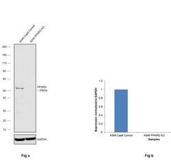
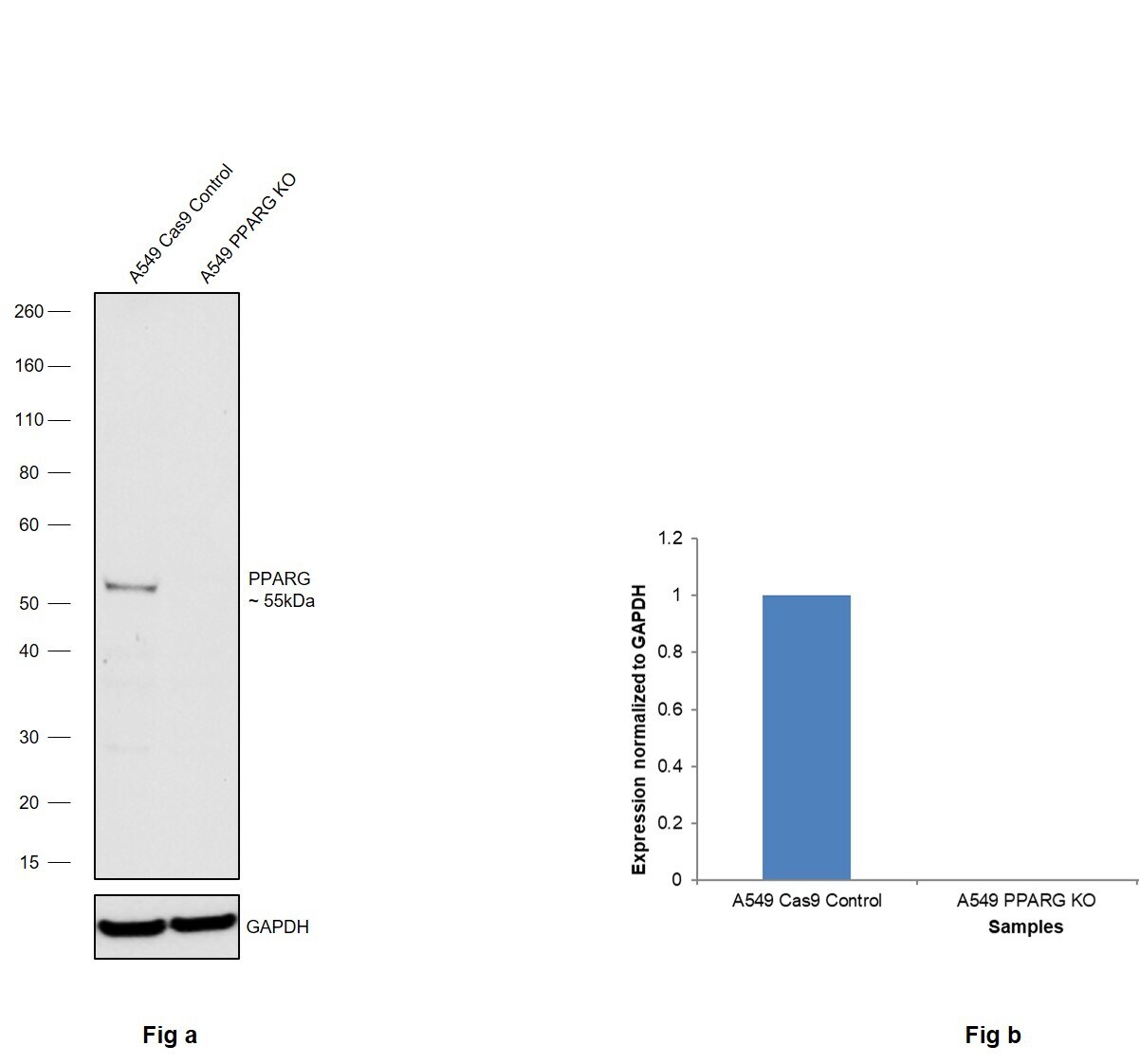
- Knockout of PPARG was achieved by CRISPR-Cas9 genome editing using LentiArray™ Lentiviral sgRNA (Product # A32042, Assay ID CRISPR751810_LV) and LentiArray Cas9 Lentivirus (Product # A32064). Western blot analysis of PPARG was performed by loading 30 µg of A549 Cas9 (Lane 1) andA549 PPARG KO (Lane 2) whole cell extracts. The samples were electrophoresed using NuPAGE™ Novex™ 4-12% Bis-Tris Protein Gel (Product # NP0322BOX). Resolved proteins were then transferred onto a nitrocellulose membrane (Product # IB23001) by iBlot® 2 Dry Blotting System (Product # IB21001). The blot was probed with Anti-PPAR gamma Monoclonal Antibody (A3409A) (Product # 419300, 1:1,000 dilution) and Goat anti-Mouse IgG (H+L) Superclonal™ Recombinant Secondary Antibody, HRP (Product # A28177, 1:6,000 dilution) using the iBright FL 1000 (Product # A32752). Chemiluminescent detection was performed using SuperSignal™ West Dura Extended Duration Substrate (Product # 34076). Loss of signal upon CRISPR mediated knockout (KO) using the LentiArray™ CRISPR product line confirms that antibody is specific to PPARG.
Supportive validation
- Submitted by
- Invitrogen Antibodies (provider)
- Main image
- Experimental details
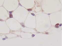
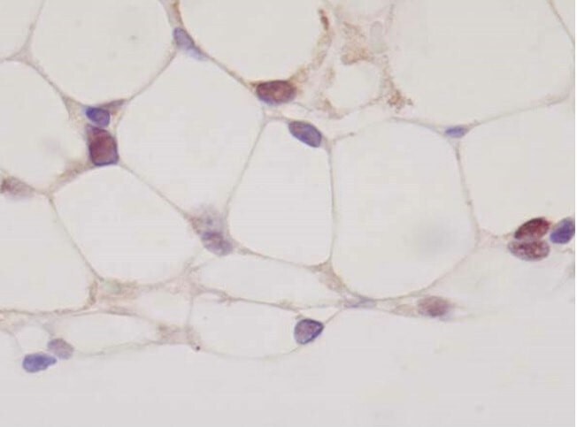
- Immunohistochemistry analysis of PPAR gamma in paraffin-embedded rat adipose tissue. The sample was incubated in a PPAR gamma monoclonal antibody (Product # 419300) at a dilution of 20 µg/mL. Section was fixed with 10% formalin-fixed in immersion, activated by autoclave at 121°C for 15 minutes, and blocked with 3% H2O2 in methanol.

- Submitted by
- Invitrogen Antibodies (provider)
- Main image
- Experimental details
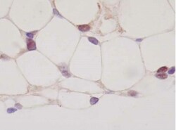
- Immunohistochemistry analysis of PPAR gamma in paraffin-embedded rat adipose tissue. The sample was incubated in a PPAR gamma monoclonal antibody (Product # 419300) at a dilution of 20 µg/mL. Section was fixed with 10% formalin-fixed in immersion, activated by autoclave at 121°C for 15 minutes, and blocked with 3% H2O2 in methanol.
Supportive validation
- Submitted by
- Invitrogen Antibodies (provider)
- Main image
- Experimental details
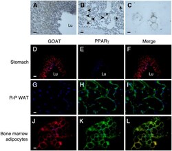
- Figure 3 Tibial marrow adipocytes express GOAT. Visualization of GOAT expression using DAB ( A - C ) and fluorescence ( D - L ) IHC in stomach ( A , D - F ), retroperitoneal WAT ( G - I ) tibial bone marrow ( B ) and lipid-containing tibial bone marrow cells ( C , J - L ). In DAB images GOAT expression is shown in brown (Arrowheads ( B ) show GOAT-positive adipocytes and arrows show adjacent smaller cells). Fluorescent images ( D - L ) show nuclei stained with DAPI (blue), GOAT expression in red ( D , G , J ), PPARgamma expression in green ( E , H , K ) and co-expression of GOAT and PPARgamma in yellow in merged images ( F , I , L ). Scale bar: 50 mum ( A , B , D - I ), 25 mum ( C , J - L ); Lu stomach lumen.
